

“A patient can have as many diseases as he or she pleases.” Many (if not most) of us have heard some variation on this axiom. It’s a little poetic, quite memorable, and 100% true. Moreover, those diseases are not limited to the eye.
It’s also been said that glaucoma is “a sick eye in a sick body” because a number of conditions and treatments involving the cardiovascular, endocrine, immune, and neurologic systems can impact the incidence and progression of both primary open-angle glaucoma (POAG) and primary angle-closure glaucoma (PACG).1 This article will provide a high level (and certainly not exhaustive) review of the prevailing wisdom around systemic diseases and medications that increase or decrease the risk of glaucoma.
The cornerstone of effective glaucoma risk assessment is a comprehensive understanding of the patient’s overall health status and medication profile, and particularly in the elderly, comorbidities and polypharmacy are more often the rule than the exception. Glaucoma is increasingly being recognized as a multifactorial disease that involves far more than simply elevated intraocular pressure (IOP); it is influenced by vascular, metabolic, inflammatory, and neurologic factors.2 A thorough case history identifies relevant conditions and medications, and informs the ophthalmic investigation that follows.3
Primary Open-Angle Glaucoma
Systemic Blood Pressure and Ocular Perfusion Pressure
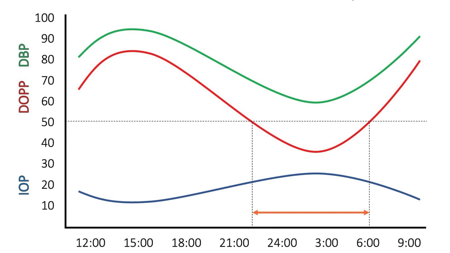
Although elevated blood pressure (BP) is known to lead to a modest increase in IOP, its impact on the incidence of POAG appears to be negligible.4 However, systemic hypotension, particularly iatrogenic low BP resulting from the aggressive treatment of systemic hypertension, has long been recognized as contributing to both the onset and progression of glaucomatous optic neuropathy (GON).5 An arguably overly simplified but still helpful clinical construct to inform risk assessment is diastolic ocular perfusion pressure (DOPP), the difference between diastolic BP (DBP) and IOP (DOPP=DBP-IOP).6 Note that both a decrease in DBP and an increase in IOP, a combination that occurs frequently during the nocturnal period, can compromise DOPP (Figure 1). A number of large epidemiologic and longitudinal studies have identified an association between low DOPP and the incidence, prevalence, and progression of POAG across multiple diverse populations.5,7,8 More specifically, these and other investigations have suggested that a DOPP in the low 50s in an individual treated for systemic hypertension significantly increases the risk of glaucoma onset and progression.9 The evolution of GON in the face of low in-office IOPs likely warrants 24-hour ambulatory blood pressure assessment to ascertain the contribution of DOPP.
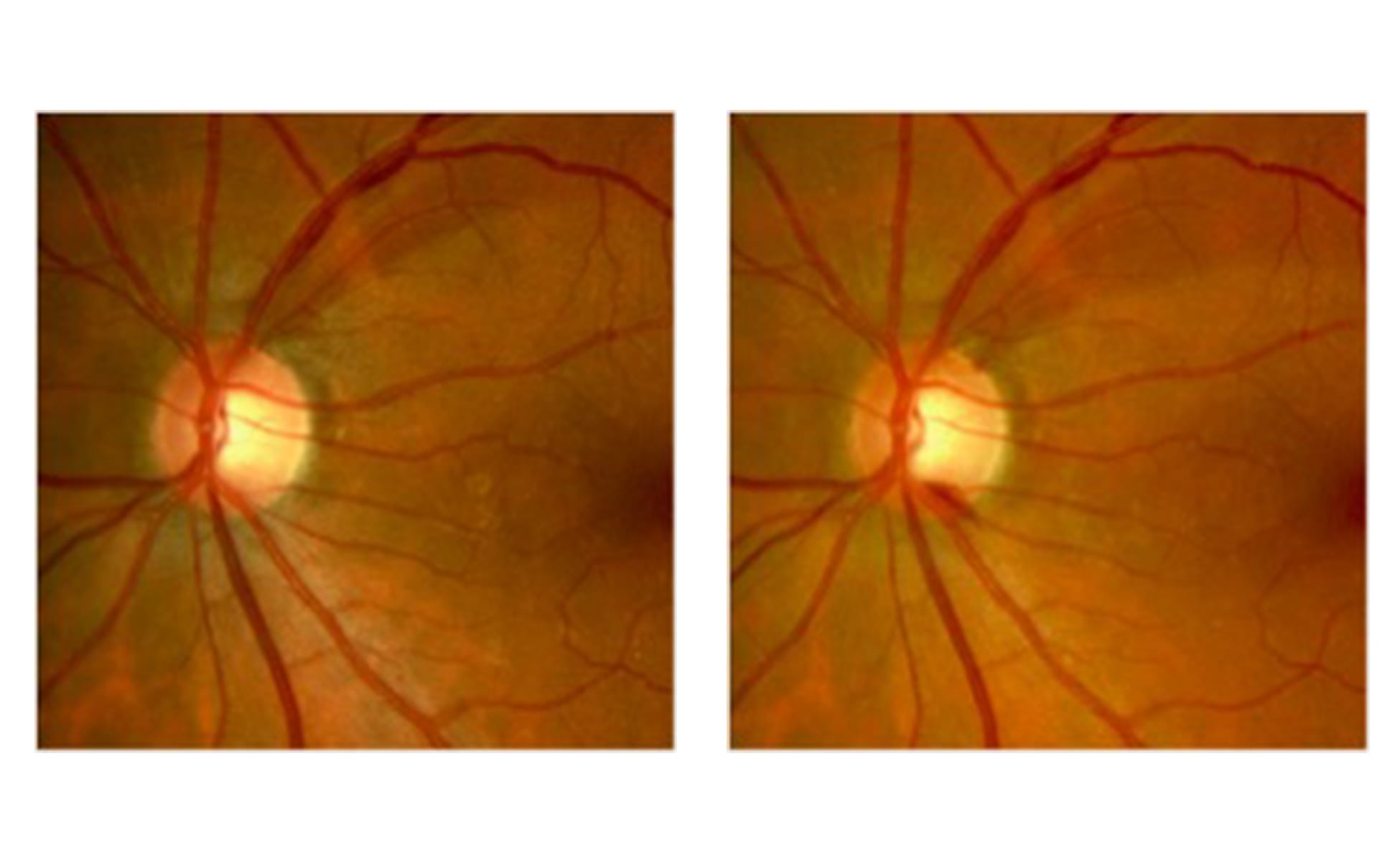
More recently, it has come to light that the type of antihypertensive medication also appears to matter. In prospective investigation, review of insurance data, and cross-sectional analyses, the use of systemic calcium channel blockers (CCBs) was associated with a nearly 40% greater odds of POAG (Figure 2).10 In juxtaposition to earlier hypotheses, rather than being neuroprotective (through addressing inappropriate vasospasm and faulty autoregulation in cases of primary vascular dysregulation characterized by migraine, Raynaud phenomenon, and decreased sensation of thirst), CCBs may in fact be “directly detrimental to retinal tissue.”11
In contrast, the use of diuretics, angiotensin receptor blockers, angiotensin-converting enzyme inhibitors, or systemic beta blockers (BBs) was not associated with increased risk of glaucoma.12 In fact, systemic BBs were protective due to their impact on IOP, but clinicians should also be cognizant of the fact that the impact of a topical BB may be blunted in patients who already use the medication systemically. In at-risk patients using CCBs for hypertension, angina, or arrhythmia, consulting their primary care practitioner (PCP) about the feasibility of alternate treatments could (some would argue should) be considered.
The timing of treatment for high BP may also be contributory. A marked drop in nocturnal BP, regardless of the type of drug used, exacerbates retinal nerve fiber layer and visual field loss.13,14 Again in consultation with the PCP, consideration may be given to using antihypertensive medications in the morning, with the goal of maintaining a normal nocturnal physiologic BP dip of 10% to 15%, while avoiding iatrogenic overdipping and the possibility of hypoxic end-organ damage.15
Diabetes
Diabetes mellitus has long been investigated as a risk factor for POAG. Although this was initially somewhat controversial, the contemporary understanding is that chronic hyperglycemia contributes to microvascular dysfunction, neuroretinopathy, oxidative stress, and prolonged inflammation, all of which may increase retinal ganglion cell (RGC) susceptibility.16 Interestingly, however, the treatment of diabetes appears to reduce the risk of glaucoma to a greater degree than would be expected based upon its impact on glycemic control.
A common feature of neurodegenerative diseases, including glaucoma, is dendrite retraction and synaptic disconnection following axonal injury.17 The use of insulin, both topically and systemically, promotes regeneration of dendrites, and restores synaptic connection and RGC function, which suggests that insulin may be both neuroprotective and neurogenerative.18 In fact, in light of these data, some have gone as far as to call POAG “type 4 diabetes”; others have previously termed Alzheimer disease “type 3 diabetes.”19,20
The use of metformin is associated with a dose-dependent reduction in risk of POAG that is independent of both HbA1c and IOP. In a 2015 study, individuals using a daily dose of 2 mg metformin for 2 years showed a 21% reduced risk of glaucoma, whereas the Rotterdam study suggested an even more impressive odds ratio (OR) of only 0.18 for incident POAG in patients taking metformin.21,22 Metformin possesses geroprotective (antiaging) properties that target mitochondrial dysfunction, fibrosis, inflammation, ischemia, and oxidative stress, and they reduce risk and severity of age-related disease, including glaucoma.23 This raises the fascinating, albeit somewhat outlandish, question of whether age could eventually become another modifiable risk factor in the treatment of POAG.
Finally, glucagon-like peptide 1 receptor agonists (GLP-1 RAs) potentiate glucose-dependent insulin secretion and inhibit glucagon release, appetite, and gastric emptying. These drugs also cross the blood-brain and blood-retina barriers and exhibit neuroprotective properties, enhancing mitochondrial function while decreasing neuroinflammation and glial cell activation in Alzheimer disease, Parkinson disease, stroke, and POAG.24,25 Specific to the latter, GLP-1 RAs reduce the risk of incident POAG with a robust hazard ratio of 0.08 that appears to be independent of any impact on HbA1c and IOP, but may depend upon duration of treatment greater than 3 years.26
So, which is better, metformin or GLP-1 RAs?
In a recent retrospective comparison of 124,000 patients with type 2 diabetes, those treated with GLP-1 RAs showed a significantly decreased risk of POAG vs those treated with metformin (relative risk of 0.59 at 1 and 3 years), prompting the investigators to conclude that “although metformin’s neuroprotective effects are promising, the variability in its impact on glaucoma risk suggests that its benefits may be less consistent or potent compared with those of GLP-1 receptor agonists.”27
Although it is exciting to think that we are on the cusp of long-awaited IOP-independent neuroprotective glaucoma therapies, we must bear in mind that these data are based upon post hoc analyses of nonrandomized cardiovascular studies and must be corroborated by large, well-designed prospective randomized controlled trials with primary ophthalmic endpoints.28
Obstructive Sleep Apnea
Obstructive sleep apnea (OSA) is characterized by repeated episodes of partial or complete upper airway collapse during sleep, leading to recurrent hypoxia that increases the risk of diabetes, heart disease, and stroke.29 It is a very common condition with a prevalence as high as 4% in men that is estimated to affect 1 billion individuals worldwide.30 A relationship between OSA and POAG has long been suspected (primarily driven by chronic ischemia/reperfusion injury), but whether OSA was causative or simply a comorbidity remained in question until relatively recently.
In contemporary meta-analyses and narrative reviews, OSA was found to confer an OR of up to 3.6 for concurrent POAG, a risk that increases to 5.5 with more severe OSA.31 Moreover, the course of glaucomatous disease is often more severe in patients with concurrent OSA. In patients with normal-tension glaucoma (NTG; arbitrarily defined as GON in the presence of statistically normal untreated IOP), the prevalence of OSA was more than triple that in controls, whereas in patients with moderate/severe OSA, the prevalence of NTG was nearly 7 times that in those with no or mild OSA.32
Although continuous positive airway pressure (CPAP) therapy does appear to help stabilize the oxygen supply to the optic nerve head, it has also been implicated as increasing nocturnal IOP via elevated intrathoracic pressure and mechanical globe compression.33 This effect has prompted some researchers to suggest that patients with comorbid OSA and POAG should opt for treatments other than CPAP; however, this conclusion remains controversial, and requires more study before a firm consensus can be reached.34
Corticosteroids
The impact of corticosteroids on IOP, whether (in order of increasing risk) systemic, periocular, topical, or intraocular, has been recognized for decades.35 With tens of millions of prescriptions written annually, the likelihood of a patient in your chair having a history of steroid use is quite high.36 Exposure to steroids increases resistance to aqueous outflow through the trabecular meshwork by suppressing phagocytosis, stiffening actin cross-linking, and increasing extracellular matrix material, potentially leading to ocular hypertension and secondary OAG.37 As many as 1 in 3 individuals are “steroid responders,” and 1 in 20 experience an IOP increase of more than 15 mmHg or to a level of greater than 31 mmHg (Figure 3).38 Individuals who have high myopia, POAG, or a family history of POAG are at increased risk.39 IOP usually normalizes within weeks of steroid discontinuation, but can become and remain quite elevated with chronic steroid use. Moreover, the ophthalmic consequences of steroid use persist, and can create diagnostic uncertainty in a patient with GON and statistically normal IOPs. In other words, is this potentially progressing NTG, or remote steroid induced glaucomatous damage that is unlikely to change over time?

The Importance and Challenge of Adherence
As the common adage posits, “Drugs don’t work for patients who don’t take them,” and yet 50% of patients treated for glaucoma discontinue treatment within 6 months, mirroring statistics for other chronic diseases.40 Treatment of an initially asymptomatic disease must be convenient, affordable, and tolerable. Poor adherence, whether triggered by medication burden (both topical and systemic), adverse effects, costs, or a lack of appreciation for the serious nature of a disease, sets the stage for potentially preventable progression.41 Indeed, in the Collaborative Initial Glaucoma Treatment Study, patients who reported never missing a dose of medication lost -0.62 dB mean deviation over 8 years of follow-up, while those who missed doses at two-thirds of visits lost -2.23 dB.42 An educated and engaged patient is more likely to be an adherent patient, and an adherent patient is less likely to experience unnecessary functional loss. Glaucoma, like life, is a team sport.
Primary Angle-Closure Glaucoma
As a backdrop, PACG, although less common than POAG, particularly in North America, is responsible for a disproportionate amount (~50%) of glaucoma-related vision loss worldwide.43 Multiple nonmodifiable risk factors for angle-closure disease have been identified, including East Asian ethnicity, advancing age, female sex, hyperopia, and a family history of PACG.44,45 However, the impact of systemic medications is significant, and cannot be overlooked.
Drug-Induced Acute Primary Angle Closure
Many commonly prescribed systemic medications can precipitate acute primary angle closure (APAC, a true ocular emergency) through either pharmacologic mydriasis inducing pupillary block or ciliochoroidal effusion triggering ciliary block.46 The 3 drugs with the highest OR for APAC are the antimigraine agent sumatriptan (OR 12.6), the antiepileptic topiramate (OR 5.1), and the serotonin norepinephrine reuptake inhibitor duloxetine (OR 4.0).47 The mechanism of action for the latter is felt to be mydriasis induced by anticholinergic and adrenergic effects that cause pupillary block.48 Fortunately, discontinuing duloxetine; initiating standard treatment with topical miotics, aqueous suppressants, and steroids; and performing a therapeutic laser peripheral iridotomy (LPI) are usually successful in breaking the angle closure attack.
Both sumatriptan and topiramate, however, can cause bilateral APAC through idiosyncratic ciliochoroidal effusion, which leads to anterior displacement of the lens/iris diaphragm and ciliary block.49,50 This can occur within weeks, and is accompanied by an often dramatic myopic shift.51 Unlike pupillary block, ciliary block should not be managed with oral carbonic anhydrase inhibitors, pilocarpine, or LPI, all of which can exacerbate the anterior lens/iris movement and prolong the attack.52 Rather, treatment entails immediate drug discontinuation and the use of topical steroids to quell inflammation and cycloplegic agents (both rapidly acting phenylephrine and longer acting cyclopentolate) to counter the anterior shift of the lens and iris.
For this reason, differentiating pupillary from ciliary block is critical. The latter often presents with uniform flattening of the entire anterior chamber, significant myopic shift, and a history of contributory medication use or recent ophthalmic surgery.53
It has been estimated that 1 in every 3 cases of APAC arise at least in part due to the influence of systemic medications.54 With more than 60 drugs known to increase the risk of APAC, and more than 40 million prescriptions written in North America each year for duloxetine, sumatriptan, and topiramate alone, failure to identify susceptible individuals and precipitating medications is a missed opportunity in the prevention of APAC.
Conclusion
In many, if not most, of our patients who have or are at risk of glaucoma, systemic comorbidities and polypharmacy are the norm. An awareness of the intersection of general and ocular health, directed by a current and detailed case history for the individual in question, arms the clinician with the information needed to provide personalized care and partner with both the patient and their multidisciplinary health care team to set the stage for optimal outcomes. OM
References
-
Pache M, Flammer J. A sick eye in a sick body? Systemic findings in patients with primary open-angle glaucoma. Surv Ophthalmol. 2006;51(3):179-212. doi:10.1016/j.survophthal.2006.02.008
-
Alqawlaq S, Flanagan JG, Sivak JM. All roads lead to glaucoma: induced retinal injury cascades contribute to a common neurodegenerative outcome. Exp Eye Res. 2019;183:88-97. doi:10.1016/j.exer.2018.11.005
-
Coleman AL, Miglior S. Risk factors for glaucoma onset and progression. Surv Ophthalmol. 2008;53 Supple1:S3-10. doi:10.1016/j.survophthal.2008.08.006
-
Leske MC. Ocular perfusion pressure and glaucoma: clinical trials and epidemiologic findings. Curr Opin Ophthalmol. 2009;20(2):73-78. doi:10.1097/ICU.0b013e32831eef82
-
Memarzadeh F, Mei Y-L, Chung J, Azen SP, Varma R, Los Angeles Latino Eye Study Group. Blood pressure, perfusion pressure, and open-angle glaucoma: The Los Angeles Latino Eye Study. Invest Ophthalmol Vis Sci. 2010;51(6):2872-2877. doi:10.1167/iovs.08-2956
-
Costa VP, Harris A, Anderson D, et al. Ocular perfusion pressure in glaucoma. Acta Ophthalmol. 2014;92(4):e252-e266. doi:10.1111/aos.12298
-
Tielsch JM, Katz J, Sommer A, Quigley HA, Javitt JC. Hypertension, perfusion pressure, and primary open-angle glaucoma. A population-based assessment. Arch Ophthalmol. 1995;113(2):216-221. doi:10.1001/archopht.1995.01100020100038
-
Hulsman CAA, Vingerling JR, Hofman A, Witteman JCM, de Jong PTVM. Blood pressure, arterial stiffness, and open-angle glaucoma: the Rotterdam Study. Arch Ophthalmol. 2007;125:805-812. doi:10.1001/archopht.125.6.805
-
De Moraes CG, Cioffi GA, Weinreb RN, Liebmann JM. New recommendations for the treatment of systemic hypertension and their potential implications for glaucoma management. J Glaucoma. 2018;27(7):567-571. doi:10.1097/IJG.0000000000000981
-
Vergroesen JE, Schuster AK, Stuart KV, et al. Association of systemic medication use with glaucoma and intraocular pressure: the European Eye Epidemiology Consortium. Ophthalmology. 2023;130(9):893-906. doi:10.1016/j.ophtha.2023.05.001
-
Kastner A, Stuart KV, Montesano G, et al. Calcium channel blocker use and associated glaucoma and related traits among UK Biobank participants. JAMA Ophthalmol. 2023;141(10):956-964. doi:10.1001/jamaophthalmol.2023.3877
-
Leung G, Grant A, Garas AN, Li G, Freeman EE. A systematic review and meta-analysis of systemic antihypertensive medications with intraocular pressure and glaucoma. Am J Ophthalmol. 2023;255:7-17. doi:10.1016/j.ajo.2023.03.014
-
Krasińska B, Karolczak-Kulesza M, Krasiński Z, et al. A marked fall in nocturnal blood pressure is associated with the stage of primary open-angle glaucoma in patients with arterial hypertension. Blood Press. 2011;20(3):171-181. doi:10.3109/08037051.2010.538964
-
Melgarejo JD, Lee JH, Petitto M, et al. Glaucomatous optic neuropathy associated with nocturnal dip in blood pressure: findings from the Maracaibo Aging Study. Ophthalmology. 2018;125(6):807-814. doi:10.1016/j.ophtha.2017.11.029
-
Charlson ME, de Moraes CG, Link A, et al. Nocturnal systemic hypotension increases the risk of glaucoma progression. Ophthalmology. 2014;121(10):2004-2012. doi:10.1016/j.ophtha.2014.04.016
-
Song BJ, Aiello LP, Pasquale LR. Presence and risk factors for glaucoma in patients with diabetes. Curr Diab Rep. 2016;16(12):124. doi:10.1007/s11892-016-0815-6
-
Di Polo A. Insulin for retinal ganglion cell regeneration in glaucoma. Invest Ophthalmol Vis Sci. 2023;64(8):2404.
-
Agostinone J, Alarcon-Martinez L, Gamlin C, Yu W-Q, Wong ROL, Di Polo A. Insulin signalling promotes dendrite and synapse regeneration and restores circuit function after axonal injury. Brain. 2018;141(7):1963-1980. doi:10.1093/brain/awy142
-
Dada T. Is glaucoma a neurodegeneration caused by central insulin resistance: diabetes type 4? J Curr GlaucomaPract. 2017;11(3):77-79. doi:10.5005/jp-journals-10028-1228
-
de la Monte SM, Wands JR. Alzheimer's disease is type 3 diabetes - evidence reviewed. J Diabetes Sci Technol. 2008;2(6):1101-1113. doi:10.1177/193229680800200619
-
Lin H-C, Stein JD, Nan B, et al. Association of geroprotective effects of metformin and risk of open-angle glaucoma in persons with diabetes mellitus. JAMA Ophthalmol. 2015;133(8):915-923. doi:10.1001/jamaophthalmol.2015.1440
-
Vergroesen JE, Thee EF, Ahmadizar F, et al. Association of diabetes medication with open-angle glaucoma, age-related macular degeneration, and cataract in the Rotterdam Study. JAMA Ophthalmol. 2022;140(7):674-681. doi:10.1001/jamaophthalmol.2022.1435
-
Daka Q, Neziri B, Lindner E, Blanco AA. Metformin in glaucoma treatment. J Glaucoma. 2024;33(6):387-393. doi:10.1097/IJG.0000000000002387
-
Mouhammad ZA, Vohra R, Horwitz A, et al. Glucagon-like peptide 1 receptor agonists - potential game changers in the treatment of glaucoma? Front Neurosci. 2022;16:824054. doi:10.3389/fnins.2022.824054
-
Sterling JK, Adetunji MO, Guttha S, et al. GLP-1 receptor agonist NLY01 reduces retinal inflammation and neuron death secondary to ocular hypertension. Cell Rep. 2020;33(5):108271. doi:10.1016/j.celrep.2020.108271
-
Sterling J, Hua P, Dunaief JL, Cui QN, VanderBeek BL. Glucagon-like peptide 1 receptor agonist use is associated with reduced risk for glaucoma. Br J Ophthalmol. 2023;107(2):215-220. doi:10.1136/bjophthalmol-2021-319232
-
Muayad J, Loya A, Hussain ZS, et al. Comparative effects of glucagon-like peptide 1 receptor agonists and metformin on glaucoma risk in patients with type 2 diabetes. Ophthalmology. 2025;132(3):271-279. doi:10.1016/j.ophtha.2024.08.023
-
Luo Y, Xia Y, Gong X, Hao M, Wei Q, Liao L. GLP-1 receptor agonists in eye disease: a comprehensive review of current research and future potential. BMC Ophthalmol. 2026;26(1):12. doi:10.1186/s12886-025-04559-x
-
Mannarino MR, Di Filippo F, Pirro M. Obstructive sleep apnea syndrome. Eur J Intern Med. 2012;23(7):586-593. doi:10.1016/j.ejim.2012.05.013
-
Benjafield AV, Ayas NT, Eastwood PR, et al. Estimation of the global prevalence and burden of obstructive sleep apnoea: a literature-based analysis. Lancet Respir Med 2019;7(8):687-698. doi:10.1016/S2213-2600(19)30198-5
-
Liu S, Lin Y, Liu X. Meta-analysis of association of obstructive sleep apnea with glaucoma. J Glaucoma. 2016;25(1):1-7. doi:10.1097/IJG.0000000000000357
-
Bilgin G. Normal-tension glaucoma and obstructive sleep apnea syndrome: a prospective study. BMC Ophthalmol. 2014;14:27. doi:10.1186/1471-2415-14-27
-
Kiekens S, De Groot V, Coeckelbergh T, et al. Continuous positive airway pressure therapy is associated with an increase in intraocular pressure in obstructive sleep apnea. Invest Ophthalmol Vis Sci. 2008;49(3):934-940. doi:10.1167/iovs.06-1418
-
Chen H-Y, Chang Y-C, Lin C-C, Sung F-C, Chen W-C. Obstructive sleep apnea patients having surgery are less associated with glaucoma. J Ophthalmol. 2014;2014:838912. doi:10.1155/2014/838912
-
Bartlett JD, Woolley TW, Adams CM. Identification of high intraocular pressure responders to topical ophthalmic corticosteroids. J Ocul Pharmacol. 1993;9(1):35-45. doi:10.1089/jop.1993.9.35
-
Waljee AK, Rogers MAM, Lin P, et al. Short term use of oral corticosteroids and related harms among adults in the United States: population based cohort study. BMJ. 2017;357:j1415. doi:10.1136/bmj.j1415
-
Clark AF, Wordinger RJ. The role of steroids in outflow resistance. Exp Eye Res. 2009;88(4):752-759. doi:10.1016/j.exer.2008.10.004
-
Tripathi RC, Tripathi BJ, Haggerty C. Drug-induced glaucomas: mechanism and management. Drug Saf. 2003;26(11):749-767. doi:10.2165/00002018-200326110-00002
-
Mitchell P, Cumming RG, Mackey DA. Inhaled corticosteroids, family history, and risk of glaucoma. Ophthalmology. 1999;106(12):2301-2306. doi:10.1016/S0161-6420(99)90530-4
-
Cramer JA. Effect of partial compliance on cardiovascular medication effectiveness. Heart. 2002;88(2):203-206. doi:10.1136/heart.88.2.203
-
Harasymowycz P, Birt C, Gooi P, et al. Medical management of glaucoma in the 21st century from a Canadian perspective. J Ophthalmol. 2016;6509809. doi:10.1155/2016/6509809
-
Newman-Casey PA, Niziol LM, Gillespie BW, Janz NK, Lichter PR, Musch DC. The association between medication adherence and visual field progression in the Collaborative Initial Glaucoma Treatment Study. Ophthalmology. 2020;127(4):477-483. doi:10.1016/j.ophtha.2019.10.022
-
Quigley HA. The number of people with glaucoma worldwide. Br J Ophthalmol. 1996;80(5):389-393. doi:10.1136/bjo.80.5.389
-
Sun X, Dai Y, Chen Y, et al. Primary angle closure glaucoma: what we know and what we don’t know. Prog Retin Eye Res. 2017;57:26-45. doi:10.1016/j.preteyeres.2016.12.003
-
Xu BY, Friedman DS, Foster PJ, et al. Ocular biometric risk factors for progression of primary angle closure disease: the Zhongshan Angle Closure Prevention Trial. Ophthalmology. 2022;129(3):267-275. doi:10.1016/j.ophtha.2021.10.003
-
Lachkar Y, Bouassida W. Drug-induced acute angle closure glaucoma. Curr Opin Ophthalmol. 2007;18(2):129-133. doi:10.1097/ICU.0b013e32808738d5
-
Na KI, Park SP. Association of drugs with acute angle closure. JAMA Ophthalmol. 2022;140 (11):1055-1063. doi:10.1001/jamaophthalmol.2022.3723
-
Wu A, Khawaja AP, Pasquale LR, Stein JD. A review of systemic medications that may modulate the risk of glaucoma. Eye (Lond). 2020;34(1):12-28. doi:10.1038/s41433-019-0603-z
-
Abtahi M-A, Abtahi S-H, Fazel F, et al. Topiramate and the vision: a systematic review. Clin Ophthalmol. 2012;6:117-131. doi:10.2147/OPTH.S27695
-
Hsu C-R, Chen Y-H, Tai M-C, Lu D-W. Sumatriptan-induced angle-closure glaucoma: a case report. Medicine (Baltimore). 2017;96(22):e6953. doi:10.1097/MD.0000000000006953
-
Lee JTL, Skalicky SE, Lin M-L. Drug-induced myopia and bilateral angle closure secondary to zolmitriptan. J Glaucoma. 2017;26(10):954-956. doi:10.1097/IJG.0000000000000742
-
Murphy RM, Bakir B, O’Brien C, Wiggs JL, Pasquale LR. Drug-induced bilateral secondary angle-closure glaucoma: a literature synthesis. J Glaucoma. 2016;25(2):e99-105. doi:10.1097/IJG.0000000000000270
-
Quigley HA, Friedman DS, Congdon NG. Possible mechanisms of primary angle-closure and malignant glaucoma. J Glaucoma. 2003;12(2):167-180. doi:10.1097/00061198-200304000-00013
-
Wu AM, Stein JD, Shah M. Potentially missed opportunities in prevention of acute angle-closure crisis. JAMA Ophthalmol. 2022;140(6):598-603. doi:10.1001/jamaophthalmol.2022.1231