Malpractice
Management
It's Good to
be Suspicious
This unusual case stresses the importance of glaucoma screening.
 by Jerry Sherman,
O.D., F.A.A.O. with
Jeffrey Roth, O.D. and Herminder Boparai, O.D.
What's the likelihood of a scientist, who has devoted much of his career to studying glaucoma in rats, suffering substantive vision loss from undetected glaucoma while under the care of a world-famous glaucoma specialist? And what's the likelihood that this Ph.D. would work alongside yours truly and that I would examine him in an international endeavor to prevent blindness from another optic neuropathy? As my friend Lou Catania, O.D., often says, "It ain't rare if it's in your chair!"
Looking out for a colleague
Dr. P (for patient) wound up in my chair in Brazil during the first week of October 2003. We were both part of an interna-tional research team evaluating the world's largest Leber's Hereditary Optic Neuropathy (LHON) pedigree. Because Dr. P was wearing thick minus lenses, I wanted to screen him for a retinal nerve fiber layer (RNFL) defect. (I believe and teach that all myopes have glaucoma until proven otherwise.) One year earlier in Brazil, I meant to screen Dr. P for this disease, but I unfortunately never had the opportunity.
During a break when we both had several minutes to spare, I used a new scanning laser polarimeter with variable cornea compensator on Dr. P. This instrument has a screening mode that's capable of evaluating both eyes of a typical patient in about one minute.

|

|

|

|
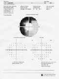
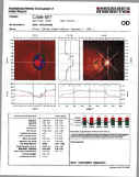
| Diagnostic synergy OD. Note the excellent correspondence of the RNFL loss measured by scanning laser polarimeter with variable cornea compensator and the central and peripheral fields. Also note how the field defect extends from the central field to the peripheral field. | |||
Cause for concern
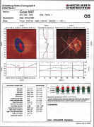
At the time, the only relevant information that I had was from my observations that Dr. P was about 50 years of age and was close to a 10.00D myope. The polarimeter results were grossly abnormal in each eye. The nerve fiber indicator (NFI) was 71 OD and 57 OS (both abnormal).
The NFI is a computer-generated neural net number that represents the artificial intelligence of a computer that has been trained to distinguish normal RNFL patterns from glaucomatous RNFL patterns. The NFI runs from a low of two to a high of 98. An NFI higher than 30 occurs in more than 90% of glaucoma patients and an NFL lower than 30 occurs in more than 90% of normal patients.
In addition to the abnormal NFI, many of the temporal, superior, nasal, inferior, temporal (TSNIT) parameters were abnormal as shown by the color-coded probability symbols (see Figure on page 24 and below). The deviation map revealed focal areas of RNFL reduction, also depicted by color-coded probability symbols.
Next, I measured Dr. P's Goldmann IOPs at 18 mmHg OU and performed confocal scanning laser imaging to assess the disc topography. In contrast to the scanning laser polarimeter results, the disc parameters as measured by the confocal scanning laser were all within normal limits. Ophthalmoscopy was difficult through an undilated pupil, but I thought the discs were tilted with a large but shallow cup in each eye. Both eyes demonstrated a mild posterior staphyloma, which is a common finding in a high myope.
Not wanting to alarm Dr. P, I suggested that we again measure his IOPs and obtain visual fields early the next morning, before the LHON subjects arrived.
Following the clues
Dr. P mentioned that a prominent, university-based, glaucoma specialist, Dr. D (for doctor) back in the United States was following him annually. According to the specialist, Dr. P's pressures were always borderline and his last visual fields, a 24-2 obtained less than one year earlier, were normal.
Curiously, Dr. P mentioned that his daughter and wife thought that he was more tentative when driving and that he moved his eyes and head quite a bit more while in the car. Dr. P interpreted that change in driving behavior as likely a result of worsening of his peripheral visual field. Based on this information, I made a mental note to obtain both a 30-degree and 30-60-degree visual field the next morning.

|
 |
 |

|
|
Diagnostic synergy OS. Note the excellent correspondence of the RNFL loss measured by scanning laser polarimeter with variable cornea compensator and the central and peripheral fields. Also note how the field defect extends from the central field to the peripheral field. |
|||
Facing the diagnosis
Considering the abnormal scanning laser polarimeter and the subjective complaints, it was an easy prediction that the peripheral fields would be abnormal in both eyes.
In a recently completed but unpublished study, we now have evidence that the scanning laser polarimeter has a much higher correlation with the entire field (0-60 degrees) than to the central 24-2. This makes sense because the scanning laser polarimeter measures all the axons within the RNFL and not just that small number of axons that correspond to the central 24 degrees.
|
|
|
|
Scanning laser polarimeter with variable cornea compensator testing revealed significant RNFL loss OD > OS.
NFI: OD=71, OS=57. |
|
The 8:00 a.m. IOPs the next morning were 23 mmHg OU. The visual field tests revealed loss within the central 30 degrees in each eye and the loss corresponded to the field defects found with the 30-60 field test.
Contrasting the central and peripheral fields, it's obvious that the peripheral field loss is more profound than the central loss, especially in the left eye. Because the 24-degree fields were normal less than one year earlier and Dr. P has had symptoms of peripheral field loss for more than one year, he and I conclude that he had lost a large chunk of his peripheral fields in each eye while retaining a completely normal central field. We now have evidence of this in many other glaucoma patients.
A number of glaucoma specialists worldwide rely on the 24-2 degree central field and on the laser scanning tomography for disc topography. Dr. P still has a normal optic nerve head tomography analysis and had a normal central fields less than one year earlier.
However, both the scanning laser polarimeter with variable cornea compensator and the peripheral fields show a profound loss. Because the scanning laser polarimeter RNFL loss corresponds and even predicts the central and peripheral field loss, the synergy of these findings increases their diagnostic power. Such synergy, defined when the whole is greater than the sum of its parts, becomes irrefutable.
This synergy of structure and function may well be the future of early glaucoma detection.
An inevitable end
Will Dr. P sue Dr. D for missing the glaucomatous damage that was most likely present less than one year earlier? Most patients in a similar situation wouldn't, but Dr. P is so knowledgeable about glaucoma -- and its likely progression -- that he certainly might. He has published all about glutamate toxicity and the cascading of neurotoxins in eyes that have moderately advanced glaucoma.
Dr. P knows from his rat studies that once glaucomatous damage gets to a certain stage, there's no guarantee that lowering the pressures will halt the damage. Because Dr. P already has symptoms associated with his loss of visual field, he recognizes that his field loss will likely only progress.
Gaining insight
Dr. P learned a lot about glaucoma in humans during the 10 days he spent in Brazil. Although a Ph.D. and not a clinician, he now recommends a peripheral field and a scanning laser polarimeter with variable cornea compensator for all glaucoma suspects to other professionals in the field. So do I.


