pachymetry
Measuring Up with Pachymetry
This diagnostic tool can change
patients' lives. See why you shouldn't practice without it.
BY THOMAS MILLER, O.D., F.A.A.O., Fayetteville, N.C.
|
|
|
|
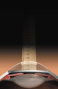
ILLUSTRATION BY SHARON AND JOEL
HARRIS |
|
As we're pressured to keep up with technology's mind-boggling pace, it's tempting to discount the need for some of the more expensive ophthalmic instruments. However, some are necessary because they give us information that we can't obtain by any other means. The corneal pachymeter is one of these vital instruments. It's cost-effective and should be available to any practitioner who manages glaucoma, co-manages refractive surgery or fits contact lenses.
Over the past few years, much has been written about the role of corneal pachymetry in eye care. Since I obtained my pachymeter more than three years ago, I have become a firm believer in its usefulness on many fronts. That's why I'd like to speak to the many optometrists who are sitting on the fence: Those of you who are trying to determine why you should add this technology to your practices and those of you who are convinced that you don't need it.
Abundant applications
It's common knowledge that the two main uses of corneal pachymetry in the optometric practice is for glaucoma diagnosis and refractive surgery comanagement. We could even argue that involvement in either of these endeavors without measuring corneal thickness is paramount to substandard practice.
But the benefits of pachymetry aren't limited to glaucoma and LASIK. It's also useful in monitoring conditions of the cornea such as corneal edema, Fuch's dystrophy, bullous keratopathy, posterior polymorphous dystrophy, contact lens overwear, herpes keratitis and keratoconus. I have also noted some usefulness in monitoring traumatic corneal edema and it's resolution over time. This may provide an explanation for reduced acuity in some of these cases.
Diagnosing glaucoma
As more of us diagnose and treat patients who have glaucoma, it's imperative that we do so with a planned purpose. Researchers have predicted a substantial rise in the number of people who have glaucoma in the next 20 years and we need to use every instrument at our disposal to properly treat these individuals.
The clinical usefulness of pachymetry has been proven through years of research and practice. Cases have shown diagnosed glaucoma patients who
have been able to discontinue medications after finding that they actually had thick corneas, which artificially raised their IOPs. Alternatively, many documented cases demonstrate that patients exist who have thinner-than-normal corneas with artificially lowered pressures. (This can sometimes provide us with an explanation for continued glaucomatous cupping under seemingly normal pressures.)
Selecting surgical candidates
It's well known that refractive surgery procedures depend heavily on healthy corneas. Therefore, we must evaluate patients who have thinner-than-normal corneas more critically. Sometimes a patient's corneal thickness will be the deciding factor in whether she's a good candidate for refractive surgery or it can indicate what type of surgery may best benefit her.
Take, for example, the case of a 34-year-old pediatrician interested in LASIK. Her habitual prescription was 3.00 1.50 x 170 OD and 2.50 1.00 x 163 OS. Her exam, including a cycloplegic refraction and corneal topography, was unremarkable. Pachymetry measured at 460 µm, roughly equal in each eye. In consultation with a refractive surgeon, we decided that she wasn't a good candidate for LASIK based on thin corneas, so this patient didn't waste a trip to the surgery center. It's also beneficial to know your patient's corneal thickness after she's undergone refractive surgery so you can calculate her true IOP.
Getting a patient's real IOP
The reason for performing pachymetry is to establish a more realistic IOP based on a patient's corneal thickness. You may have two patients, each with pressures of 25 mmHg and similar disc changes. If one has a corneal thickness of 480 µm and the other is 580 µm, then their target pressures will be completely different because their corrected pressures are probably eight to 10 points apart, instead of being the same at 25 mmHg.
It's also been hypothesized that a thin cornea may correlate to having a thinner-than-normal scleral, and thus a thin, weakened lamina cribosa at the optic nerve head. This would make sense. We already know that those with thin corneas are predisposed to developing glaucoma. Perhaps a weakened lamina cribosa is the deciding factor and the reason that some develop damage at lower pressure than others. Possible correlation of low central corneal thickness with biochemical changes in scleral collagen or systemic parameters awaits further investigation.
CASE STUDY #1. A 53-year-old African-American male who had 0.70./0.70 cupping and IOPs of 19 mmHg in each eye and with non-discrete, but suspicious visual field changes. Conventional thinking might say this patient is a glaucoma suspect based on the cupping, repeat pressures and fields. Corneal pachymetry showed this gentleman has central corneal thickness (CCT) of 440 µm in each eye. Assuming a popular and widely accepted conversion factor of 2.5 mmHg for every 50 µm of change from normal (»?550 µm) would give this patient a true IOP reading of 24 mmHg. Certainly enough of a difference to cause me to rethink his treatment and perhaps start pressure-lowering medication.
CASE STUDY #2. A 60-year-old female who has repeat IOPs of 25 mmHg and with 0.40/0.40 cupping and clean visual fields. We could classify her as a glaucoma suspect or perhaps an ocular hypertensive. Pachymetry readings show her CCT to be thick, measuring at 680 µm in each eye. A quick conversion showed me that her true pressures were closer to 19 mmHg.
Without a pachymetric measurement, both patients in these case studies would have suffered, either by not getting necessary treatment or by continuing to take expensive medication unnecessarily.
|
Third-Party Payers & Pachymetry |
|
Most major medical plans consider ultrasound corneal pachymetry medically necessary for the following indications:
Reimbursement ranges from about $19 from Medicare to $50 from some private medical plans. A technician can perform the procedure itself in about two minutes. |
|
Coding and billing pearls
The determination of corneal thickness has historically been coded with 0025T. Recently, however, this code has changed. 0025T is the old category-III CPT code and is defined as "determination of corneal thickness (e.g., pachymetry) with interpretation and report, bilateral." Unfortunately, we can only measure each patient's corneal thickness once in a lifetime.
76514 Ophthalmic ultrasound, echography, diagnostic; corneal pachymetry, unilateral or bilateral (determination of corneal thickness) is the new CPT code. However, some payers are slow to adapt to the new code and still accept or demand the older 0025T code. It does seem to be a source of confusion, real or imaginary, to some of them.
Do the right thing
Advances in technology have benefited doctors and patients alike. We'd all like to add a host of excellent instruments to our practices, but a corneal pachymeter may be one of those that, 20 years from now, we'll wonder how we ever practiced without it. As with many other advanced instruments at our disposal, the pachymeter isn't a one-test diagnostic instrument. Instead, it's simply another device that we have in our optometric toolboxes.
I typically explain pachymetry to patients as the difference between pressing on a basketball and pressing on a balloon (on the premise that the exterior of the ball is thicker than the balloon). This helps them understand the concept of thin corneas giving different pressure readings than thick ones. For the cost of a few thousand dollars, you can gain peace of mind, knowing that you've done all that's possible for your patients. Do them a favor before submitting them to a lifetime of expensive treatment. Give them the benefit of the doubt before forever changing their lives with the psychologically devastating diagnosis of an incurable, potentially blinding disease.
References available on request.
|
Correction Values |
|
| CORNEAL THICKNESS (MICRONS) | CORRECTION FACTOR |
| 405 | 7 |
| 425 | 6 |
| 445 | 5 |
| 465 | 4 |
| 485 | 3 |
| 505 | 2 |
| 545 | 0 |
| 565 | -1 |
| 585 | -2 |
| 605 | -3 |
| 625 | -4 |
| 645 | -5 |
| 665 | -6 |
| 685 | -7 |
| 705 | -8 |


