contact lens
Bandage Contact Lenses to the Rescue
Soft contact lenses greatly increase our success in
treating corneal abrasions.

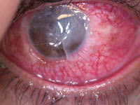
It's Monday morning and three cases of corneal abrasion or corneal pathology present to your office. Or you're co-managing refractive surgery and a patient returns one day postoperatively with PRK (photorefractive keratectomy) rather than LASIK (laser-assisted in situ keratomileusis). What do you do?
Dealing with abrasions

Other challenges include significant photophobia, sharp eye pain and blepharo-spasm. In addition, these patients are anxious because they don't see well. Doctors know that this condition is self-limiting and frequently doesn't require any medical treatment, with resolution occurring in 24 to 48 hours.
But abrasion patients don't always experience pain in the same way. Some are stoic and have minimal complaints. Others are extremely unhappy. Therefore, treating pain can be as important as resolving the epithelial defect in abrasion management. Bandage soft contact lenses (BSCL) address all the concerns inherent in corneal abrasions.
Introducing the BSCL
|
BSCLs at Your Disposal |
|
PureVision, Bausch & Lomb Focus Night & Day, CIBA Vision ProClear, Coopervision Acuvue Advance, J & J Bandage Lens, Optik K & R CLPL Simplon Therapeutic, UltraVision |
For years, the first choice of treatment for corneal abrasions was the pressure patch (PP). When appropriately applied, the PP prevented excessive eye movement, accidental aggravation or another injury to the eye. It also kept the eye closed. It was theorized also that the eye was continually bathed in antibiotic solution or ointment to minimize the chance of microbial keratitis. Although helpful, the PP didn't seem to provide pain relief, with patients often needing narcotic analgesics.
Two events changed abrasion management. The first was a series of reports that the PP did not improve corneal healing over eyes without the PP. The second was the introduction of the therapeutic or bandage soft contact lens (BSCL) in the mid 1970s.
With the PP seemingly irrelevant in healing the cornea, practitioners re-examined its relative inability to reduce pain substantially and prevent repeat injury. The BSCL met these two criteria and even one-upped the opaque PP, allowing the patient to see out of the injured eye instead of obscuring vision.
|
Fitting Pearls |
|
Other tips I've picked-up on using BSCLs to treat
corneal abrasions include the following:
► "I tend
to use silicone hydrogels with the lowest BC [base
curve] ... to minimize lens movement on the eye for
traumatic corneal abrasions or lacerations up to 3mm to
4mm average diameters," says Dr. Jan Boehringer, an
optometrist in Indiana.
|
Since its inception, the BSCL seemed like the perfect solution for the abrasion. Initial patient acceptance was very high because pain relief was almost immediate and was dramatic. But the early BSCLs had problems. Their low water content (normally in the FDA Group 1 Lens Class), may have simplified handling, but it also interfered with normal corneal physiology because they were too thick or got dirty too quickly. And the lenses were expensive, sometimes costing $50 each.
However, these early lenses worked. In one of my first cases in the late 1970s, a young woman who injured her eye when she reached for a large can of tomato sauce on a high shelf was referred to my office. The can fell against her eye, causing an 8mm penetrating wound. Postoperatively, she experienced intolerable ocular pain and was refused analgesics because of her past history of difficulty in metabolizing them. I tried a FDA-labeled Group 1 BSCL and it immediately relieved her pain. I was impressed! But what troubled me was the relatively poor physiologic performance of these lenses. In this case, the cornea swelled sufficiently for me to see endothelial folds.
Just a couple of years after the Group 1 lenses were introduced, Group 4 55% water content soft contact lenses arrived and helped somewhat. Comfort was unquestionably better. One of the first Group 4 lenses I used, the Hydrocurve II (CIBA Vision), seemed to work well when worn up to 48 hours continuously, but tended to dehydrate faster than the low-water content, Group 1 lenses. Thus, they ultimately caused even more discomfort than the Group 1 lens. However, their larger size did help in covering more of the corneas and seemed to move less on an eye without excessive topographical irregularity.
BSCLs in everyday practice
|
|
|
Case 1: A fingernail caused this abrasion. |
Now we'll examine how BSCLs can help with the abrasion problems that can walk into your office any day. The following three examples are real-life cases I've had.
■ CASE 1. A 35-year-old woman accidentally scratched her own eye with her fingernail as she slept. She presented to the emergency room the next morning with pain she rated as 10 out of 10 (in other words, debilitating), photophobia and 20/200 vision in the affected eye. The abrasion was 6mm x 5mm (horizontal x vertical) and centered on the visual axis. If I had seen this patient 20 years earlier, I would have immediately instilled copious amounts of antibiotic ointment and applied a PP. My emergency room coverage over the years made me quite skilled at this.
I started the patient on erythromycin ophthalmic ointment five to six times a day to minimize lid interaction on the cornea and antibiosis. I then added cyclopentolate 1% four times a day, to control the pain. While pain decreased after one hour, it was never eliminated until I added hydrocodone (Vicodin, Abbot), one tablet, every six hours.
The abrasion resolved without the use of the BSCL, but there were significant pain issues that required prescription analgesia. With this classical management technique, successful and complete resolution of the abrasion is possible with relevant antibiosis and appropriate oral analgesia. But the patient still experiences pain and discomfort.
|
|
|
Case 2: Central corneal abrasion upon presentation. |
■ CASE 2. A 50-ish woman felt something in her eye while cleaning and vacuuming her house. She irrigated her eye extensively and thought that she had removed the foreign body. Three days later, she experienced intense (9/10 or 10/10) eye pain, photophobia and 20/100 vision. The photograph below shows oblong, 6mm high x 5mm wide corneal abrasion, vertically oriented. In this case, the mechanism of the abrasion was different from that of the patient in our first case, although similar in that an initial foreign body caused the injury and its unsophisticated removal probably caused the abrasion. I inserted a Group 4 lens with a 8.9mm base curve and 15mm diameter. The patient felt better immediately.
What made this especially unique is her historical use of a fentanyl transdermal patch (Duragesic, Johnson & Johnson) for unremitting back pain. With this kind of systemic analgesia, any additional analgesia for the relief of eye pain would have been minimal.
The tip from this case is the concomitant use of antibiotics with a BSCL in place. Doctors have dipped the lens in antibiotic solution or applied it topically over the lens while in situ or before lens placement. We don't know for certain whether one method of antibiotic support for a lens in situ is better than the other. What we do know is that the antibiotic delivery vehicle can destabilize the lens fit. Despite the destabilization, some form of antibiotic support is necessary. In my opinion, I think the use of topical ophthalmic solution may be better than ointment.
When the patient applied the erythromycin ointment, the lens immediately moved excessively on blink. I believe that the ointment increased, rather than minimized, the friction between the lens and the lid. In the end, this patient healed nicely with a return to 20/25 vision and a very small scar.
|
|
|
Case 3: Corneal laceration with bandage lens situ. |
■ CASE 3. This one is seemingly complicated. It illustrates the principles of corneal wound care. The patient was a 42-year-old male who was assaulted and experienced a ruptured globe including multiple fractures of the face, nose and orbit. At day one post-op he received numerous corneal sutures to close a corneal laceration wound. The next day, he appeared for follow-up with a loosely applied patch over the eye. He had loosened the patch throughout the night in order to instill topical medications.
With a history of an open wound, endophthalmitis became the most significant potential post-operative complication. I knew that if I were to use a BSCL on this patient, it must not increase or enhance the prospect for endophthalmitis. I ended up using a large-diameter, Group 4 hydrogel lens. This lens, though, moved off-center on each blink when out of the bottle. The photo above (top) shows the lens not only has a bubble underneath, but is also de-centered. Repeated lens dislodgement occurred until the fifth reinsertion finally was stable on the eye.
Expanded applications
In these three cases, the traditional crop of bandage lenses seemed adequate for continuous wear of up to seven days. To expand the repertoire of conditions for BSCL use to include recurrent corneal erosion, bullous keratopathy and severe corneal autoimmune conditions, a better performing lens was necessary. The ideal BSCL should have excellent or superior oxygen transmissibility, be easy to handle and stable on the eye. The silicone hydrogel (SiHy) lens seems to be the answer for many of the current clinical and performance concerns.
|
|
| Case 3: Initial presentation status post-op with corneal sutures in place. |
Low in water content, these lenses handle much like the traditional Group 1 lens. However, they exceed the oxygen transmissibility of current high-Dk, hydrogel lenses.
In fact, I've heard many leading O.D.s say that the silicone hydrogel lens is their first choice BSCL. Dr. Angelo De Vivo, a Mentor, Ohio optometrist with broad experience in refractive surgery co-management and corneal disease, gives high praise to the material as a BSCL. "Today, the SiHy [BSCL] is my first choice over traditional patching," he says. "I use these [BSCLs] for keratitis, corneal abrasions and PRK postoperative management, as well as RCE [recurrent corneal erosion]."
Ease their pain
Corneal abrasion management is no longer just about managing wound closure. It is also about pain management. With the appropriate armamentarium of BSCLs, oral analgesics, topical and oral antibiotics, excellent results are possible. Whether you use the traditional hydrogel or the newer silicone hydrogel materials, the BSCL will provide satisfactory results if fit for complete corneal coverage and centered with minimal movement.
So, when you see that next 4mm x 4mm corneal abrasion, grabbing the BSCL rather than that oral narcotic and PP may be your best plan of action.
Dr. Hom is Coordinator, Primary Care Optometry, San Mateo Medical Center. Reach him through his web site at http://www.geocities.com/rchom/


