Ditch the Itch
A variety of medications are at your
disposal to help assuage your allergy patients' symptoms.


The price of managing the 50 million Americans who suffer from allergy may cost up to $18 billion, estimates the Asthma and Allergy Foundation of Amer-ica. As the allergy season approaches, you must remain aware of these options so that your patients have the best opportunity for relief of the typical and, in some cases, complex complications of ocular allergy.
Immunology
Ocular allergy is the result of either
a type-I hypersensitivity (anaphylactic) response or a type-IV, cell-mediated immune
complex response. Type-I reactions are caused by exposure to an antigen that's either
airborne, food-born, insect-born or by drugs. These reactions typically occur within minutes of exposure to
the offending item. Along with ocular tissue, the skin and tissues of the respiratory
tract and gastrointestinal tract may be affected. Clinically, the local responses
of the aforementioned tissue results in conjunctivitis, asthma, hives and eczema.
Allergen activation of B-lymphocytes initiates the cascade of events that results in an immediate (anaphylactic), hypersensitivity reaction. The first step is the production of the immunoglobulin IgE. IgE is present primarily in the skin and mucous membranes and is a critical player in type-I allergic reactions.
Mast cells and basophils are leukocytes (white blood cells) present in blood and tissue that become sensitized as IgE binds to their surface. When exposure
to the allergen recurs, mast cell membrane permeability is altered, allowing calcium ions to invade the cell. This causes a process known as degranulation. Degranulation allows the contents held within the cell to escape. When phospholipase A2, one of the contents within the mast cell, is liberated it contributes to the break down of other membrane phospholipids into arachidonic acid.
The cyclooxygenase pathway converts the arachidonic acid into prostaglandins, prostacyclin and thromboxane A2. The lipox- ygenase pathway converts these chemicals to leukotrienes. Histamine, a preformed mediator is also released. Via both H1 and H2 receptors, histamine plays a key role in type-I reactions. As a result of these chemicals stimulating nociceptors and by histamine stimulation of H1 re- ceptors, an increase in vascular permeability and neural triggering inspires itching and the contraction of smooth muscle within the respiratory and digestive tract. The stimulation of H2 receptors and exposure to chemical cytokines results in vasodilatation, itching, mucous production and an increase in gastrointestinal secretions. These pathways produce red, itchy eyes, rhinitis and gastrointestinal upset.

The delayed hypersensitivity reaction or the type-IV reaction involves T-lymphocytes (or T cells). The type-IV response can be caused by various offending agents including viruses, bacteria, fungi, rejection of graft tissue, tumors, chemical exposure, and in cases of contact dermatitis, a variety of contact substances such as plants, deter- gents, and latex rubber.
In response to exposure to an offending agent, antigen-presenting cells introduce antigen to the T cells, resulting in their activation. Following the initial exposure, the process of sensitization takes place during the next one to two weeks. When the T cell is again exposed to the antigen, cytokine release occurs, activating macrophages. This results in a cytotoxic response via elevated phagocytic activity and the release of lytic enzymes. The typical delayed hypersensitivity re- sponse takes 24 hours with the worst physiologic changes occurring at 48 to 72 hours after exposure. The scenarios in which resistance occurs: The body fights the response, causing up-regulation of other chemicals and tissue constituents, eventually producing a chronic, delayed hypersensitivity reaction with the end result being fibrosis and granuloma formation.
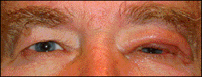
The symptoms produced by type-IV reactions pertinent to ocular allergy include contact dermatitis (see figure 4) and giant papillary conjunctivitis via the Jones-Mote reaction. As this action peaks during the course of 48 to 72 hours, the T cells and macrophages spread to the epidermis in their attempt to eliminate the antigen.
Role of the ocular surface
The anatomy of the ocular surface provides a defensive barrier as well as a potential avenue for antigens. While the pre-corneal tear film has protective value, eliminating debris from the ocular surface via blinking and flushing, it also offers allergens a media into which they may dissolve and gain exposure to ocular surface tissue. The conjunctiva provides immunity by acting as an environmental barrier, limiting the access of antigens to deeper ocular tissue. The conjunctiva also provides a specific immune response, as it contains T- and B-lymphocytes.
Classification
For some, a detailed work-up including tear film assay, conjunctival biopsy, skin testing or consult by an allergist to specifically identify the offending agent may be beneficial, as sub categories of ocular allergy exist.
The most common sub-form of conjunctival allergy: seasonal allergic conjunctivitis (SAC) or hay fever conjunctivitis. As season's change, airborne allergens, such as pollen, become more prevalent. As these allergens come into contact with the conjunctiva, they cause SAC. Although it's less common, peren- nial allergic conjunctivitis (PAC) is caused by ubiquitous allergens, such as dust mites,
that can be found throughout the year. Classic examples of PAC allergens: pet dander, dust mites or molds. Both SAC and PAC have no pre-dilection to gender and affect patients of all ages.
Although sufferers of SAC and PAC can present with additional signs and symptoms, such as nasal allergy (rhinitis), frequently their signs and symptoms are limited to the eyes. The usual symptoms include the bi-nocular presentation of mild to moderate itching, burning, foreign body sensation and a watery or string-like mucoid discharge. Clinical signs may include a hyperemic and chemotic bulbar conjunctiva, a mild to moderate papillary reaction of the palpebral conjunctiva (see figure 1) and in chronic cases, a follicular reaction of the palpebral conjunctiva. In addition, you may observe eyelid swelling and dark pigmentation of the eyelid, due to periocular venous congestion. Corneal involvement is rare.
Vernal keratoconjunctivitis (VKC) is much less common than either SAC or PAC. It classically emerges in the spring and summer months. Sufferers are most commonly male children, between the ages of eight and 12 years old. Because the condition is seasonal, it's considered to be a self-limited problem. VKC usually declines in severity in the late teen years and may stop its chronic, periodic cycle by the patient's early 20's.
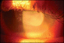
Similar to SAC and PAC, VKC produces bulbar and palpebral conjunctival signs, however, those associated with VKC are typically more severe than the other sub categories. When coagulated exudate adheres to the inflamed palpebral conjunctiva, a pseudomembrane is formed. These membranes loosely adhere to the conjunctiva and can be removed by peeling with forceps or a moist cotton swab. Along with allergic conjunctivitis, pseudo-membranes may be found in cases of epidemic keratoconjunctivitis (EKC), ligneous conjunctivitis and bacterial infections. In addition, corneal findings are common with VKC by way of a mechanism produced by the irregular superior conjunctiva eroding the conjunctiva. Corneal signs may range from punctate epithelial keratopathy to areas of corneal epithelial macroerosion and shield ulceration (see figure 3, above).

The etiologies of all ocular allergic
reactions are sufficiently similar in that the resulting therapeutic managements
are comparable, with only slight variation. Management is primarily aimed at reducing
the patient's symptoms and when necessary, preventing ocular tissue damage through
inflammation control. The type and frequency of medications depends on the diagnosis,
severity of the condition and your management style.
Topical supportive thera-pies. One function of the biological tear film: to wash
away de- bris and environmental allergens from the ocular surface. The mechanical
flushing action produced by the eyelids is capable of removing allergens and other
waste, reducing or even canceling the allergic response. Considering that many patients
with allergic symptoms have dry eye as a primary symptom, you must rule out tear
deficiency in chronic, mild allergic conditions. Clinical examination may include
the measurement of the tear break up time, quantity of the tear prism and lacrimal
lake and Schirmer testing to aid in the assessment of the tear film.
For patients with dry-eye-related
burning, gritty sensation or irritated eyes, artificial tears (preserved and unpreserved
varieties) may suffice. For the relief of simple itching, remove the offending agent,
and prescribe a soothing cold compress and simple topical antihistamine preparations,
such as emadastine difumerate (Emadine, Alcon) and levocabastine hydrochloride (Livostin,
Novartis) at a q.i.d. dose, which should be adequate.
Antihistamine/mast cell stabilizers.
Although the combination of topical mast cell stabi- lizers and antihistamines is
the newest category of anti-allergy medications, they are the medications-of-choice
when treating ocular
allergy. This is because mast cell stabilizers work by stabilizing the receptors
on mast cell vesicles before they can degranulate, preventing allergen/mast cell
coupling, thus inhibiting degranulation and cytokine expression (this begins the
cycle of the allergic response). This blocking of the histamine/H1 receptor interaction
provides relief from histamine activity.

In December of 1996, the first medication developed in this category was approved for use in the United States. That medication, olopatadine hydrochloride 0.1% (Patanol, Alcon) remains successful today. The recommended b.i.d. dosing is convenient and provides many patients with the ability to continue contact lens wear during allergy season. The launch of the newest formulation, Pataday (0.2% olopatadine hydrochloride) allows for qd dosing.
Ketotifen fumerate (Zaditor, Novartis),
azelastine hydrochloride (Optivar, Bausch & Lomb) and epinastine (Elestat, Allergan)
work by a similar mechanism and are effective as b.i.d.-use medications. In October
of 2006, Novartis announced the U.S. Food and Drug Administration's (FDA) approval
of Zaditor, in its current formulation, as an over-the-counter product made available
January, 2007. olopatadine hydrochloride
0.1%, ketotifen fumerate and epinastine are available in 5-mL
bottles, while azelastine hydrochloride is supplied in a 6-mL bottle. This drug
is marketed with b.i.d. dosing, and the 6-mL bottle typically provides 50 days of
treatment.
Topical steroids. In episodes
in which acute signs and symptoms become present or in severe cases of allergy,
topical steroids are an option. These agents decapitate the inflammatory cascade,
inhibiting the conversion of phospholipase A2 to arachidonic acid. Loteprednol etabonate
0.2% suspension (Alrex, Bausch & Lomb) is effective and can be used qd to q.i.d.
for management of ocular inflammation. When you desire more immediate relief of
acute ocular symptoms associated with severe ocular inflammation, we recommend lote-
prednol etabonate 0.5%, (Lote-max, Bausch & Lomb).
Topical antibiotics have no role in the treatment of ocular allergy, making combination prep-arations (topical antibiotic/ste- roidal agents) a poor choice. In fact, adding more medication into the mix of an individual encountering an allergic response increases the possibility of producing a corneal toxic response or medicamentosa keratitis. However, when tissue changes associated with the allergic response are responsible for breaching the corneal epithelium, as in cases of keratopathy or a shield ulcer, topical antibiotics may be required to prevent infection of the open cornea.
Other topical medications. Some mast-cell stabilizing medications require q.i.d. dosing. These include pemirolast potassium 0.1%, (Alamast, Santen Pharmaceuticals), lodoxamide tromethamine 0.1% (Alomide, Alcon), cromolyn sodium 4% (Crolom, Bausch & Lomb) and cromolyn sodium 4% (Opticrom, Allergan). Nedocromil sodium 2% (Alocril, Allergan), another alternative, is typically used b.i.d.
The longstanding success of the combination allergy medications and periodic supplementation with topical steroids has made the addition of the topical non-steroidal anti-inflammatory class (NSAID) rare. However, in those instances when topical ste-roids are either contraindicated or the allergic response has approached the most severe levels, these agents are available and work synergistically. The NSAID ketorolac tromethamine 0.4% (Acular LS, Allergan) provides excellent relief of ocular surface pain and discomfort and is typically administered on a b.i.d. to q.i.d. dosing schedule.
An additional medication that has
been used successfully in clinical trials to reduce the symptoms and signs of VKC:
topical cyclosporine. Concentrations of 1.0%, 1.25% and 2.0% topical cyclosporine
have been used
without major side effects and provide relief from this form of chronic allergy. Ocular allergy, as a producer of
signs and symptoms, has a misleading reputation. Minimalists often use the
diagnosis to connote temporary, nebulous symptomology, occurring in the absence
of obvious infection. However, from the patient�s perspective, the inconvenience
and chronic nature of the problem produces frustration and annoyance. Clearly
medications can help these individuals restore homeostasis and increase their
quality of life. So, be sure to explore all the options at your disposal.
References: available upon request.


