contact lens management
"Eccentricity" is in Against Thin
GP lens provides keratoconus patients with comfort and stable visual acuity.


DIANNE ANDERSON, O.D., F.A.A.O. AND RANDY KOJIMA, F.O.A.A.
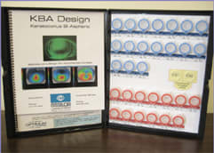
The KBA (keratoconus bi-aspheric) lens, from Essilor Contact Lens, enables keratoconic patients to achieve optimal centration, peripheral alignment, comfort and acuity.
The KBA is a 10.2mm diameter GP design with a 9.2mm back surface optic zone. The large optic zone combined with a custom aspheric back surface allows for centration over the pupil and, thus, reduced flare and glare. Also, the aspheric front surface eliminates the radial astigmatism created by the highly aspheric back surface to provide quality visual acuity (VA). Further, KBA comes with software to help you determine parameters.
Fitting philosophy
The KBA is designed to protect the corneal epithelium from GP-lens-induced abrasions and keratoconus-caused scarring. All KBA lenses exhibit clearance over the cone's apex, as lens touch or bearing on the cone increases corneal scarring risk. Also, the KBA enables you to match its eccentricity (e-value) to that of the cornea.
Corneas that flatten at a significant rate from their geometric center outward have high e-values, and those that flatten mildly from the geometric center have low e-values. Central nipple cones and oval cones generally have high e-values and, therefore, achieve success with the KBA's customized back-surface eccentricity. The result: optimal alignment, movement and comfort. (Note: Most topographers measure corneal eccentricity. Otherwise, you can derive the eccentricity by taking the square root of the shape factor value, e2).
Fitting methods
You can fit the KBA via:

KBA Lens MATERIAL Optimum Comfort DK: 65 WEARING SCHEDULE: Daily wear RECOMMENDED REPLACEMENT SCHEDULE: Yearly DIAMETER: 10.2mm BASE CURVE: Custom SPHERE POWER: Custom FITTING SET: 30 lenses with KBA Software included PRACTITIONER COST: $90 per warranted lens |
► Keratometry. Select the base curve of the initial diagnostic KBA by subtracting 0.6mm from the flat central corneal radius in millimeters and using the 0.98e KBA trial lens of equal radius.
► Topography. Determine the initial diagnostic KBA's base curve by subtracting 0.6mm from the cornea's apical radius in millimeters. Since most cones are decentered away from the corneal apex, capture a topography map on the cone apex to provide a more accurate e-value across the cone. Do this by having the patient look two to three rings upward (so the cone is centered within the Placido disk display). Use the closest KBA trial lens eccentricity (0.98e or 1.3e) to the measured cone's e-value. This will ensure clearance over the cone and result in an optimal lens-to-cornea relationship.
► Medmont topography. Apply computer-generated KBA parameters over a given topography map by selecting the "KBA" or "Advanced KBA" design feature in the E300 CL designer. Then, manipulate the base curve and e-value to create the ideal tear layer profile on the simulated sodium fluorescein (NaFl) pattern, and use the KBA trial lens with the closest base curve and eccentricity (0.98e or 1.3e) to the computer-generated pattern. (Note: Use the "Advanced KBA" designer feature to create a lens that has a low central eccentricity and a high peripheral eccentricity. The low central eccentricity enables you to fit exceptionally steep corneas and produces a flatter base curve to reduce the overall lens power. The high peripheral eccentricity results in a comfortable edge profile.)
Determining parameters
To determine the best KBA diagnostic parameters:
1. Attempt ideal cone clearance. If the trial lens exhibits excessive cone clearance, select a flatter trial lens until you achieve the ideal apical clearance. If the trial lens exhibits cone touch or bearing, select a steeper trial lens until clearance.
2. Over-refract the KBA trial lens. Perform an over-refraction on the trial lens with the ideal apical clearance (20 to 30 microns of tear-film clearance on the CL designer simulated NaFl pattern). If the patient's over-refraction VA is poor, over refract the next steeper and flatter trial lens to determine whether more or less apical clearance provides better VA. (Note: Excessive apical clearance decreases VA.)
3. Determine ideal peripheral alignment. The diagnostic trial KBA should exhibit ideal edge lift at its 3 o'clock and 9 o'clock positions. While ignoring the central fit, find the trial lens that has the best edge appearance. If the trial lens has too much edge lift, select a steeper one. If it has insufficient edge lift, select a flatter trial lens.
Enter the trial lens that has the best peripheral alignment and the trial lens that has the best apical clearance into the KBA software to view the custom base curve and eccentricity.
Add the software's tear-layer compensation value to the over refraction to calculate the final power. (Note: Increasing the KBA lens' eccentricity requires steepening the base curve to maintain the desired apical clearance or "sag." The software helps with this.)
We've found that patients fit in the large diameter KBA experience stable VA, due to its comfort and centration. And, its aspheric front surface optics improves VA by decreasing haloes and glare. OM
DR. ANDERSON PRACTICES IN AURORA, ILL., SHE SPECIALIZES IN ORTHOKERATOLOGY, KERATOCONUS, POST-SURGICAL LENS FITS AND ANTERIOR SEGMENT DISEASE. E-MAIL HER AT DIANNE.ANDERSON@COMCAST.NET.
MR. KOJIMA IS THE DIRECTOR OF TECHNICAL AFFAIRS FOR PRECISION TECHNOLOGY SERVICES IN VANCOUVER, B.C. ALSO, HE SERVES AS A PART-TIME RESEARCH SCIENTIST AND CLINICAL INSTRUCTOR AT PACIFIC UNIVERSITY COLLEGE OF OPTOMETRY IN FOREST GROVE, OREGON. E-MAIL HIM AT RANDY@PTSOPTICS.COM.


