Cracking Down on Meibomian Gland Dysfunction
Screen for suspects, inspect and express the glands, and choose the best treatments for your patients.

By Kelly Nichols, OD, MPH, PhD
So many patients are needlessly uncomfortable, suffering with ocular surface disease that can compromise the health of their eyes. In our clinic, we're involved in many research studies for dry eye and meibomian gland dysfunction (MGD), and we're always emphasizing to students that they need to look for these problems. Of course, both MGD and dry eye are inextricably intertwined. But MGD is easy to overlook as a contributing problem, especially in the absence of obviously blocked meibomian glands.
Effective therapies are available to help your patients feel more comfortable, wear contact lenses longer, and preserve the health of their eyes; we just need to accurately diagnose MGD, treat the problem and get patients on board in the process.
Keys to diagnosis
We can't diagnose MGD in a vacuum. Once you carefully consider ocular surface disease, you realize it's connected to a network of ocular surface problems, working separately and together to contribute to the frequency and severity of signs and symptoms. A patient with dry, gritty eyes might have MGD, exacerbated by ocular allergies, computer use and changing hormone levels due to menopause or any number of other factors.
The key to effective diagnosis is to make your analysis as diverse as the network of ocular surface problems. Follow these important measures:
• Ask the right questions. Although it's certainly helpful to know potential causes or issues that run concurrent to MGD — rosacea, menopause, prolonged computer use, and so on — the best clues to the disease come from questions about symptoms. Any complaint of ocular discomfort is a valid reason to suspect ocular surface disease, but some specific complaints are common among people with MGD.
Questions should delve into these areas and give patients a choice to answer yes, no or unsure:
• redness
• itching, burning
• dry eyes
• gritty feeling
• sensation that there's something in the eye
• sensitivity to eye makeup
• stopped wearing contact lenses
• sometimes need to remove contact lenses
• wearing contact lenses fewer hours per day
• eyes feel/look bad in the morning
• eyes feel/look bad in the evening
• eyes always feel bad.
If you only have time to ask 2 or 3 questions, focus on the following: 1) Do your eyes feel dry or irritated often or constantly? 2) Do your eyes ever burn or sting? If yes for 1 or 2, is it worse in the morning or at the end of a day? The doctor or technician also should ask if over-the-counter drops are being used; if so what types; and are the drops providing relief.
Any number of yes answers warrant further investigation. If patients experience burning and irritation in the morning, the problem may be anterior blepharitis and/or MGD, since dry eye alone tends to feel better in the morning and worsen as the day wears on. Patients with MGD often report mild ocular itch, which of course may also indicate allergic conjunctivitis (another problem often treated concurrently with MGD).
• Look for the signs. Because patients may have a combination of ocular surface problems, the examination helps put a name to their complaints. Check for signs of irritation such as redness and conjunctival edema. Characterize the lids, which also may show redness and edema, as well as debris or other signs of anterior blepharitis. Blocked glands may be visible or appear inflamed when you pull down the lid to carefully examine the gland orifices. Check the appearance of the glands in the upper lid. Ideally, it's a good idea to develop a habit of always checking both lids to ensure a thorough exam is performed on each patient. This helps prevents ocular surface diseases from "slipping through the cracks."
• Express the glands. Blockages clearly point to MGD, but a patient can still have the problem even without blocked glands. That's why it's essential to express the glands. Optometrists are becoming more proficient in this process (about 10% of attendees at my recent lectures raise their hands to indicate that they do this in clinic), and use of the test will increase as MGD treatments multiply.
To express the glands, use your index finger to put pressure below the lash margin for about 10 to 15 seconds. Some use a massaging or rocking movement as well. Evaluate the expressability or ease with which you can express the glands, as well as the quality of the secretions. Thick, opaque secretions or a lack of secretions point to MGD.
• Test the tear film. When we examine the tear film quantity and concentration, we can see a clear picture of the type of ocular surface disease. The tear film osmometer (TearLab) is a good global indicator of dry eye and may be a marker for treatment or research. Alternately, you can use a Schirmer's or phenol red thread test, which measures aqueous production for evaporative dry eye diagnosis (normal is approximately greater than 10 mm wetting for both tests).
• Check for surface damage. Surface damage is a real possibility for patients with MGD, especially if they've had the problem for some time. Although corneal staining may be negative for patients with mild MGD or early diagnosis, if the patient is experiencing vision problems or you suspect any damage, fluorescein or lissamine green staining can reveal it.
• Take baseline photos. Document the patient's status with a set of baseline lid photos. If the patient has lid debris, red margins and blocked glands, you can document how treatment improves over time. This is a powerful patient management tool.
At follow-up visits during treatment, the patient should be more comfortable and signs of MGD should diminish. You might test the tear film quality again for comparison. Also, you may see an improvement in blocked glands, but you should express the glands again to see if the quality and quantity of the secretions are improving.

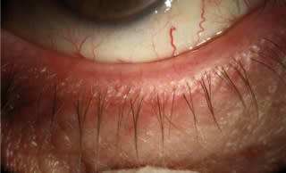
Figure 1. Mild gland plugging. Look for "capping" or white orifice openings.

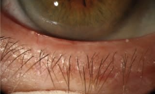
Figure 2. Moderate gland plugging. Note the increase in vasculature.

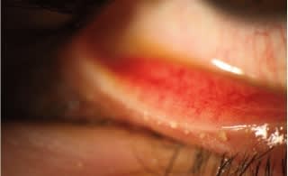
Figure 3. Meibomian gland expression before treatment. Photo courtesy of Ben Gaddie, OD, FAAO, Louisville, KY

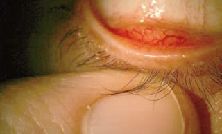
Figure 4. Meibomian gland expression after treatment. The expressibility and color have improved. Photo courtesy of Ben Gaddie, OD, FAAO, Louisville, KY
Matching patients to treatments
Initial treatment for MGD can be very subjective, since we have to determine how much a patient is willing to do on his or her own. Warm compresses and lid scrubs are the standard first steps. If they don't work, or if a patient fails to comply with recommended treatments, we can change the approach to include medication at a follow-up visit. Sometimes, at the initial visit, patients aren't willing to do this work or predict that they'll fail, in which case I might go straight to a combination of hygiene and prescribed medication.
You might choose to give patients some choices for MGD medications and supplements.
• Allergy drops: If the patient has ocular allergies, treating that problem will improve comfort and allow you to see the underlying MGD/dry eye more clearly.
• Artificial tears: Drops help soothe the ocular surface and wash debris off of the eyes and lashes. Patients may find that they need to use tears less often as treatment for MGD takes effect.
• Antibiotic ointments or drops (macrolides, aminoglycosides): Antibiotic drops/ointments are used if the patient has staph-related anterior blepharitis, although reported antiinflammatory effects of macrolides could be beneficial with inflammation due to MGD or a combination of MGD and anterior blepharitis.
• Immunosuppresive agents (cyclosporine): Cyclosporine can help improve the quantity and quality of tears for patients with aqueous deficient and mixed dry eye.
• Omega fatty acids: These supplements are thought to improve the quality of meibomian gland secretions through an anti-inflammation pathway.
• Oral tetracycline: Oral tetracyclines or doxycyclines are recommended in patients with rosacea and recalcitrant MGD.
• Steroids: Steroids may be used for acute inflammation but only for short-term treatment.
Soon, we may see standardized treatment for MGD. Because the disease is under-recognized and under-treated, the International Workshop on Meibomian Gland Dysfunction is working to develop a uniform approach. At a 2010 meeting of the Association for Research in Vision and Ophthalmology, workshop members explained that they've determined four diagnostic stages for the disease, pairing each stage with the appropriate therapeutic interventions.1
Finally, I think it's important to note that there's a frequently posed question of whether to treat asymptomatic patients with MGD. I always ask, Is the patient really asymptomatic, or did the doctor not ask the right questions? Does the patient think that his discomfort is just part of life or aging or some other contributing factor, such as allergies? Or does he just not like to complain?
MGD is not a blinding eye condition, but we don't know the long-term effects and it is a chronic disease. There could be long-term progression that we don't yet fully understand. Early diagnosis and treatment is beneficial for so many health conditions, so it's reasonable to think that it would be beneficial for MGD. I always encourage my patients to take small steps now to preserve ocular health over time, whether they have obvious symptoms or not.
Dr. Nichols is an associate professor at The Ohio State University College of Optometry in Columbus.
Reference
1. Clinicians aim to standardize meibomian gland dysfunction therapy. Ocular Surgery News U.S. Edition, June 10, 2010. Accessed online at osnsupersite.com/view.aspx?rid=64009.
Getting your message acrossWe're always working to communicate effectively with patients about their conditions. Most people have no idea that they have glands in their eyelids at all, let alone that they secrete substances that help keep their eyes moist and comfortable. Now I'm telling them that something's wrong with these glands, and they should take special lid hygiene measures, use medication and return for follow up. How do I make them understand and follow my instructions?A picture is worth a thousand wordsWhen I document the condition with some baseline photos, I show patients the red lid margins and blocked glands. I take a picture of what the secretions look like as I express the glands, too. Then I can explain in greater depth with a diagram.The visual evidence has a dramatic impact in both making patients understand their problem and motivating them to comply with treatment. It also helps to explain that treatment will improve both their comfort and the health of the ocular surface. If the patient is becoming uncomfortable in contact lenses, then I tell him that managing MGD could give him an extra few hours of comfortable wearing time each day. All of this may be new information to patients, but we can make it fairly easy to understand and treat, which means patients will be more likely to achieve positive outcomes rather than living with the uncomfortable effects of MGD. Your staff can be very influential in reinforcing your message.
Moderate turbidity (granularity of the secretions) is seen in both eyes. Photos courtesy of Katherine M. Mastrota, MS, OD, FAAO, New York, NY 
|
Optimizing Surgical OutcomesIf you're comanaging cataract patients or treating glaucoma, treating MGD before surgery may help achieve the best surgical outcomes. When a patient is measured for a premium intraocular lens, for example, the quality of the tear film on entry could affect measurement and lens selection. Patients preparing for laser vision correction should have any ocular surface problems addressed to ensure the best visual outcomes as well. What's more, a healthy ocular surface and good tear film also reduce risk for infection. |
Case Study: Classic MGDA schoolteacher learns her discomfort isn't just part of aging.GM, a 47-year-old female third-grade teacher, arrived at her 2-year eye exam and indicated on her form that she wanted to get new eyeglasses. She had been prescribed eyeglasses and contact lenses in the past, so I asked why she only wanted eyeglasses. GM replied, "I'm getting too old for contacts." As I read over the answers on her questionnaire, I could see where GM was coming from. She'd checked "yes" next to burning, itching, grittiness and redness. "It's all these years of grading papers until midnight," she said. "My eyes have had it!" I asked if her eyes bothered her in the morning, and she said they did. "That's when I used to put in my contacts — now I can't even bear the thought of putting them in." She also wrote on the form that she regularly used Visine. When I examined GM, I could see redness and slight edema of the lids and the ocular surface. I counted three blocked meibomian glands. When I expressed the glands, the secretions took about 15-20 seconds to emerge, and they appeared thick and moderately opaque. Not surprisingly, a phenol red thread test showed that GM's eyes were quite dry. I took images of both eyes, including the blocked glands and the expressed secretions. When we reviewed the images together, GM was taken aback that this problem existed without her knowledge. It was also a concern in terms of health and cosmetic appeal. I explained that her irritated eyes and inability to wear contact lenses weren't just a part of getting older. She had MGD, an uncomfortable but treatable problem. Upon seeing the images, GM was eager to begin medication and fix the problem. As a teacher, she didn't blink at being given homework. She had no hesitation in going home to start warm compresses, omega fatty acid supplements and artificial tears, and she understood that these measures could take several weeks to take effect. I saw GM again 2 weeks later. The clogged glands were gone, but all of the other signs and symptoms were identical. She said she'd been doing the compresses as ordered, but clearly they hadn't been effective. She needed further treatment. She continued the compresses, supplements, and artificial tears, and we added topical antibiotic drops once daily. When GM returned, her ocular surface and eyelids looked quieter and she had no edema. Another phenol red thread test showed that her tear volume was only slightly greater, so we went prescribed cyclosporine drops to improve her tear production and comfort. Again, we continued the hygiene and supplements. Finally, we were able to schedule the refraction visit that GM had come to me for in the first place. I was able to fit her for eyeglasses and 1-day disposable contact lenses — which she continues to wear with success. She is much more comfortable and satisfied with her eye care. From a practice management perspective, all of GM's MGD-related visits were billed medically, as were certain individual measures such as photodocumentation. TearLab osmolarity testing isn't currently reimbursable, but the company is aiming to get reimbursement for tear film osmometry in the next 18 months, barring any difficulty. What's more, because so many patients like GM exist in all of our practices, her case illustrates the enormous opportunity that MGD presents to enhance patients' health and comfort while increasing our medical billing. |


