contact lens management
Scleral Fits Simplified
With this four-step approach, you'll reduce chair time and remakes.

Gregory W. DeNaeyer, O.D.
The ability of scleral (both mini and full) lenses to vault the corneal surface makes them ideal for managing severe corneal irregularity or ocular surface disease. Some practitioners have shied away from offering scleral lenses for these conditions, however, because they believe the lens' large size will make successful fits a challenge.
Here, I dispel this belief by detailing a simple four-step approach that enables quick, successful fits. Further, following this approach will reduce chair time and lens remakes, which will improve your profit margin.
1. Select a fitting set based on lens diameter
The first step to achieving quick, successful fits with scleral lenses is to acquire a diagnostic fitting set based on the diameter of scleral lens you want to fit. Miniscleral lens diameters range from 15.0 mm to 17.9 mm and work well for mild-to-moderate corneal irregularity. Full-scleral lens diameters range from 18.0 mm to 24 mm and are capable of fitting mild-to-severe corneal irregularities, as well as managing ocular surface disease. (Note: If you're going to obtain only one diagnostic fitting set, I suggest the full-scleral lens diagnostic fitting set. Although exceptions exist, I've found that full scleral lenses don't limit you, in terms of your ability to fit one or more patients who present with severe corneal irregularities.)
2. Assess the anterior segment profile
Scleral lenses vault the cornea. Therefore, to achieve a successful fit you must be aware of the sagittal height of the anterior ocular surface. The simplest approach to this: Visually evaluate the anterior segment profile outside the slit lamp while the patient is in the exam chair, and determine whether it has a low, medium or high amount of sagittal height.
For example, an eye that has severe keratoglobus will show a relatively large sagittal height. As a result, the first diagnostic lens you apply should have a large sagittal depth.
(Note: Before applying the lens, fill it with saline to avoid air bubbles. Then, add fluorescein for diagnostic purposes. Next, instruct the patient to have his head down, and tell him to look at his feet while you apply the lens, so the fluid doesn't spill.)
3. Evaluate the fit from the center outward
Once the first diagnostic lens is in place, wait at least twenty minutes to allow it to settle. Then, evaluate the fit from the center of the lens outward. The reason: In most fitting sets, the diagnostic lenses vary by base curve with a standard haptic.
The ideal mini-scleral lens fit will have approximately 50 microns to 100 microns of corneal clearance at the highest point on the cornea, while the ideal full-scleral lens performs best with 100 microns to 200 microns of corneal clearance.
Gauge the fit by swinging your slit beam out 45° to 60° and observing the tear layer in cross section. Use white light and a thin optic section. The fluorescein in the fluid will appear bright green, allowing you to easily estimate the thickness of the tear layer. Then, compare it with the corneal thickness. Make sure the limbus is adequately cleared, as limbal bearing by the lens can damage stem cells that are imperative to corneal health.
If you note inadequate central or limbal clearance, apply a different diagnostic lens that has more sagittal depth than the first one. If the lens excessively vaults the central cornea, apply a diagnostic lens that has less depth than this lens.
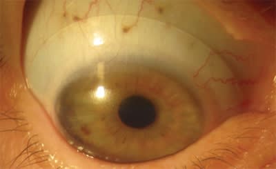
Once the lens that best vaults the cornea is in place, evaluate the haptic portion of the scleral lens. Ideally, a scleral lens should semiseal on the eye with no movement. The scleral section of the lens should rest evenly on the scleral conjunctiva without excessive bearing or impingement, which will be indicated by blanched blood vessels (See figure 1, below). Flattening the peripheral curves of the lens will remedy this situation. However, this may reduce the lens vault, resulting in corneal touch. As a result, you may need to centrally increase the lens' sagittal depth to compensate for the lens vault reduction. Steepen the peripheral curves of the lens if it's too loose or has edge lift. (Note: Some scleral lens manufacturers automatically adjust the lens' sagittal depth when flatter or steeper peripheral curves are ordered. So, check with your lab when you order the lens.)

Figure 1: Notice how the scleral section of this full-scleral lens rests evenly on the scleral conjunctiva. This is an ideal fit.
4. Over-refract
Now, its time to over-refract the best diagnostic lens. If the visual acuity with the over-refraction is less than expected or has astigmatic error, check for on-eye lens warping by performing over-keratometry of the lens, which should yield spherical readings. If the readings aren't spherical, the lens is likely bending on the eye secondary to scleral toricity. If this is the case, order a lens that is 0.2 mm thicker than the diagnostic lens to solve this problem.
If residual astigmatism occurs after the best fit is achieved, the residual correction can be provided in spectacles to be worn over the lenses. (Note: During the initial consultation when you discuss the contact lens options with the patient, it's imperative you educate him on the possibility that he may need more than one optical device to provide him with maximum vision correction. A surprised patient is often a dissatisfied patient.)
This simple, four-step approach allows you to successfully fit many irregular corneas and manage severe ocular surface disease. OM
* For trouble-shooting or guidance, seek help from the manufacturers' fitting consultant.
| Scleral Lenses |
|---|
| • DigiForm - TruForm Optics - www.tfoptics.com • Dyna Semi-Scleral - Lens Dynamics - www.lensdynamics.com • Jupiter - MedLens Innovation, LLC - www.medlens.net • Jupiter – Essilor Contact Lenses - http://essilorcontacts.com • Perimeter - Essilor Contact Lenses - http://essilorcontacts.com • Maxim - Accu Lens, Inc. - www.acculens.com • MSD (Mini Scleral Design) - Blanchard Contact Lenses, Inc. www.blanchardlab.com • So2Clear Standard - Art Optical - www.artoptical.com • So2Clear Cone - Art Optical - www.artoptical.com • So2Clear Progressive - www.artoptical.com • SoClear – Dakota Science - www.soclearlens.com |
DR. DENAEYER IS CLINICAL DIRECTOR FOR ARENA EYE SURGEONS IN COLUMBUS, OHIO AND PRESIDENT OF THE SCLERAL LENS EDUCATION SOCIETY (WWW.SCLERALLENS.ORG). E-MAIL HIM AT GDENAEYER@ARENAEYESURGEONS.COM, OR SEND COMMENTS TO OPTOMETRICMANAGEMENT@GMAIL.COM.


