


Practice pulse
TIPS, TRENDS & NEWS YOU CAN USE
ODS ON THE FRONT LINESOF FABRY DISEASE DIAGNOSIS
Be the first to identify this life-threatening disorder
Leonid Skorin, Jr., DO, OD, MS, FAAO, FAOCO
■ Optometrists are in an excellent position to identify patients who are living with a rare and inherited condition that often goes unrecognized for years. It's called Fabry disease and optometrists can be the first to diagnose it, as it presents with several ocular manifestations.
Fabry disease is an inherited disorder that primarily affects males. It's caused by the absence or faultiness of the alpha-GAL enzyme. This enzyme deficiency leads to a build up of a fatty substance known as GL-3 in cells throughout the body, which causes severe health problems, such as hypohydrosis, cerebrovascular disease and renal insufficiency or failure. Because the genetic disease is rare (roughly 10,000 U.S. cases), and its signs (angiokeratoma) and symptoms (heat and cold intolerance) vary widely, diagnosis is a challenge. (For more information on this disease, visit www.fabrycommunity.com.)
Here are the most common ocular signs of Fabry disease:
► Corneal verticillata. A total of 90% of Fabry disease patients have corneal whorling, in which dense bronze to cream-colored rays radiate from the center of the cornea.1 The corneal whorling occurs in the subepithelium, or Bowman's layer, of the cornea. A slit-lamp exam can enable you to detect the vortexshaped pattern on the cornea associated with whorling.
Patients taking antiarrhythmic drugs, such as amiodarone (Cordarone, Pfizer), may present with whorl-like opacities that mimic Fabry disease, though they, unlike Fabry disease patients, usually also experience resulting vision problems, such as glare and decreased visual acuity.
► Vascular changes on the conjunctiva and retina. Conjunctival vasculature abnormalities occur in up to 60% of Fabry disease patients, typically appearing early in the disease process.2 Specifically, the conjunctiva's small blood vessels appear sausage-like on the mucus membrane.
Retinal vascular changes associated with the condition may include arteriole constriction leading to central retinal artery occlusions, retinal and preretinal hemorrhages and hypertensive retinopathy with disc edema.3 A dilated fundus exam using high-magnification ophthalmoscopy enables you to identify these conditions.
► Cataracts. Researchers have documented two different types of lens opacities in Fabry disease: the anterior capsular, or “propeller,” cataract and the posterior subcapsular or “Fabry cataract.”4 The anterior capsular cataract develops more rarely than the Fabry cataract, and may appear as an area of translucence formed between wedged-shaped areas of cloudiness, forming the “propellerlike” pattern. The Fabry cataract typically has a dendritic-like presentation. You can best observe both cataract types via retroillumination.
If you suspect a patient has undiagnosed Fabry disease, refer him to a geneticist for confirmatory DNA testing, and encourage him to discuss the disease with his primary care doctor who can then refer him to a specialist (e.g. one who can treat one of the aforementioned associated health problems.) (See “Fabry Disease Follow-up Care” below).
By virtue of your education in eye care, you can place these patients on track to receive alpha-GAL enzyme replacement therapy—a diseasemodifying treatment which clinical research has shown may reduce the GL-3 levels in certain cells.
The result of your efforts: You'll play a role in improving the quality of life of Fabry patients, enhance your image as a vital member of the healthcare team, instill patient loyalty and attract referrals.
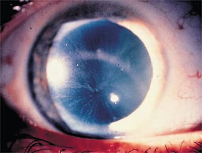
Corneal whorl in patients with Fabry disease typically has its apex below the midline of the cornea.
| Fabry Disease Follow-up Care |
|---|
| Following a confirmatory diagnosis, schedule the patient for a: • Full general ocular examination, including a visual acuity assessment • Slit lamp exam • Dilated fundus exam with direct and indirect ophthalmoscopy. Also, refer the patient to www.fabrycommunity.com or www.fabry.org to learn more about the disorder and communicate with others who have the disease. |
1. Franceshetti A. Fabry's disease: Ocular manifestations. In: Bergsma D, Bron AJ, Cotlier E (eds). The Eye and Inborn Errors in Metabolism. Vol. 12, No. 3. New York: AR Liss Co.; 1976:195-208.
2. Melton R, Thomas R. Fabry's keratopathy. Clin. Refract Optom. 2006;17:452-453.
3. Eng CM, Germain DP, Banikazemi M, et al. Fabry disease: guidelines for the evaluation and management of multi-organ system involvement. Genet Med. 2006 Sep;8(9):539-548. Review.
4. Sher NA, Letson RD, Desnick RJ. The ocular manifestations in Fabry's disease. Arch Ophthalmol. 1979:97(4):671-676.
OPTOMETRY GIVING SIGHT FUNDS INTERNATIONAL VISION CARE PROJECTS
Projects provide improved care abroad
■ Optometry Giving Sight recently provided funding for two Volunteer Optometric Services to Humanity (VOSH) International vision care projects in Central and South America. The first project will fund the purchase of equipment for an optical laboratory at the newly established School of Optometry at National Autonomous University of Nicaragua (UNAN) in Managua.
VOSH International has played an integral role in the establishment of the school, which currently has 40 students enrolled in the first year of the optometry program. The goal of the school is to educate, train and graduate local optometrists to provide affordable and accessible vision care services for the people of Nicaragua.
“We are delighted to be able to support this excellent program thanks to the funds we receive from Optometry Giving Sight,” says Larry Hookway, O.D., immediate past president of VOSH. “The school's curriculum was modeled after the La Salle program in Bogota, Colombia, and is regarded as being closest to North American educational and clinical standards.
By equipping an optical laboratory we will be ensuring that students also have access to practical training as part of their education.”
Dr. Hookway added that an important part of the education program is that students are required to provide services to poor and disadvantaged people of the country during the last 12-18 months of their curriculum.
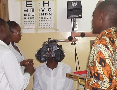
Students and faculty at the optometry school in Kumasi, Ghana with equipment provided by the International Technology Transfer program.
The second project is the VOSH International Technology Transfer Program, which supports clinical instruction by providing new and used optometric equipment to schools of optometry in Central and South America. It's anticipated that funds provided for this program will benefit optometry schools in Chile and Argentina.
Optometry Giving Sight funds sustainable eye care services for people who are blind or vision impaired due to uncorrected refractive error.
The charity works in partnership with VOSH International in the United States and has provided funds to their sustainable vision care projects in Mexico, Nicaragua, Afghanistan and Peru.
To learn more about Optometry Giving Sight, call 1-888-OGS-GIVE or visit www.givingsight.org.
Dr. Skorin is the senior staff ophthalmologist at Mayo Clinic Health System in Albert Lea, Minn.