DOCTORS RELATE THEIR EXPERIENCES WITH TOPCON’S 3D OCT-1 MAESTRO
The “Diagnostic Focus” department presents the perspectives of several optometrists regarding their experiences with one diagnostic device that has been launched recently in the United States.
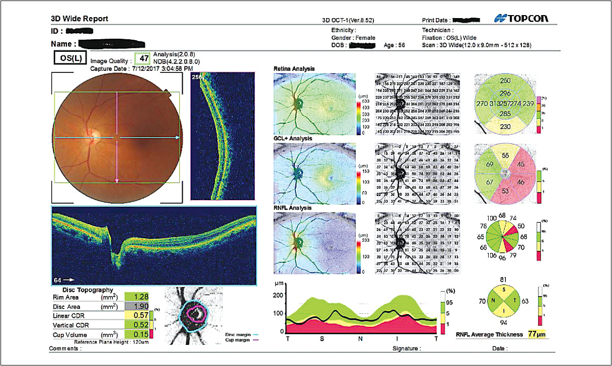
A WOMAN in her 50s with a history of multiple sclerosis presented to the office of Justin Bazan, O.D., New York, for her comprehensive eye exam. During the exam, a screening with wide-field OCT showed a dramatic thinning of her nerve fiber layer, which Dr. Bazan says he suspected was the result of optic neuritis, though not reported by the patient.
The Topcon 3D OCT-1 Maestro wide-field OCT’s scan area, which is 9mm x 12mm, facilitated the viewing of the signs of optic neuritis, specifically pallor on the optic nerve head, aiding Dr. Bazan in making the diagnosis, he says.
This column will describe how several optometrists have integrated the device into their practices, including discussions on procedure, training, practice benefits and patient care.
OVERVIEW
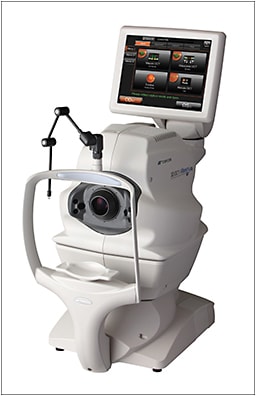
The Topcon 3D OCT-1 Maestro combines SD-OCT and a non-mydriatic fundus camera with simultaneous image capture. It offers several analysis functions, including a 12 mm x 9 mm wide-field OCT scan, anterior segment OCT, 3D macula analysis and optic disc analysis.
Physically, it measures about 46 pounds, has dimensions of about 12.1 inches to 17.4 inches wide, is 18.5 inches x 26.3 inches long and 20.4 inches to 28.4 inches high, depending on the placement of the rotatable touchscreen monitor, according to the company.
PROCEDURE
To use the device, the technician makes the appropriate touchscreen selections to coincide with the doctor’s request. Within seconds, the OCT aligns and focuses. The scan takes five to 10 seconds — at a speed of 50,000 A-scans per second, according to Topcon — before it moves to the second eye, says Jonathan Noble, O.D., Ashland, Va. In Dr. Noble’s practice, he says he uses the device when signs and symptoms indicate the need, capturing 40% to 50% of his patient base.

Travis Johnson, O.D., Murfreesboro, Tenn., says he uses the device to offer a wellness scan, estimating a capture rate of 98% of his patients. The benefits are twofold: early detection of disease states and patient education.
Specifically, Dr. Johnson says technicians walk the patient through his or her basic eye anatomy. The tech ends each patient conversation with: “The doctor is going to go over this information more carefully.” Dr. Johnson says he estimates the scan and patient education take about three to five minutes.
PRACTICE BENEFITS
Dr. Noble’s purchase of the device replaced a non-mydriatic retinal camera and retinal imaging device. The replacement allowed him to achieve a smaller footprint, he says.
Dr. Bazan says his purchase brought OCT services in-house, adding convenience for patients, increasing continuity of care and adding reimbursement possibilities. He adds that the technology reinforces his patients’ perception of his practice as state-of-the-art.

REIMBURSEMENT AND FEES
Some managed vision care plans reimburse for screening of retinal photography, but you should assess your individual contract for more details. Reimbursement for use of the device for diagnosis or monitoring disease progression can be a few hundred dollars. Please note the device does require a written interpretation and report. (For more on interpretation and report, see “Coding Strategies” on p.73.)
THE PATIENT
Dr. Bazan says he was able to use the OCT images to educate the patient about her condition.
“Patient education is enhanced when you take the time to show those images from test results,” Dr. Bazan explains. “Now, she knows. She has actually seen it and made that connection.”
He says he plans to monitor her nerve fiber layer for changes with an annual OCT scan. OM


