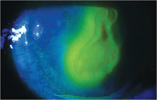
Amniotic membranes can provide relief to various corneal disease patients
Amniotic membranes can be a great treatment modality for patients who have band keratopathy, bullous keratopathy, corrosion of the cornea and conjunctival sac, corneal ulcers, keratitis and dry eye disease (DED) (moderate or severe), among other corneal issues. (See “ICD-10 Codes” for the full list of conditions.)
Here, I provide a brief overview of amniotic membranes and their insertion.

| Band Keratopathy | H18.429 |
| Bullous Keratopathy | H18.10 |
| Corrosion of Cornea and Conjunctival Sac | T26.60XA |
| Corneal Epithelial Defects | S05.00XA |
| Unspecified Corneal Ulcer | H16.009 |
| High Risk Corneal Transplants | will vary |
| In Conjunction With Superficial Keratectomy | will vary |
| Keratitis (Bacterial or Viral) | will vary |
| Pterygium | H11.009 |
| Stevens-Johnson Syndrome | L51.1 |
OVERVIEW
Amniotic membranes are harvested after planned cesarean sections and then either cryopreserved or dehydrated, comprising the two forms available to patients. For information regarding specific uses of each form, visit bit.ly/2QbKUaj (Prokera, Bio-Tissue), bit.ly/2WdOKoK (BioDOptix, Integra LifeSciences) and bit.ly/2Jofgpz (AmbioDisk, Katena).
Both types of amniotic membranes are inserted into the eye, similar to a contact lens.
Patient comfort and effica-cy remain paramount in using amniotic membranes. I find older patients and more diseased corneas have less corneal sensitivity and tolerate the tissues fairly well. I find that younger and more active patients, however, might complain of some minimal discomfort while wearing a cryopreserved amniotic membrane.
INSERTION
Historically, amniotic membranes were sutured into place to cover the cornea or the conjunc-tiva in more invasive-type procedures.
Today, however, these tissues can be kept in-office and placed in the eye by optometrists as a simple in-office procedure.
Insertion:
• Cryopreserved
A. Rinse the graft with sterile saline for a few minutes, removing the anti-infective solutions the tissue is stored in to increase patient comfort.
B. Apply a topical anesthetic to the patient’s eye.
C. Hold the superior eyelid, and have the patient look down.
D. Place the amniotic membrane underneath the superior eyelid into the superior fornix.
E. Position the amniotic membrane to make sure it’s resting under the lower eyelid for improved comfort.
F. Use a partial tape tarsorrhaphy to improve comfort.
G. Instruct the patient to move his head when looking around, rather than rolling his eye around, to help comfort.
H. Schedule the patient for a follow-up visit in three to four days (my preferred time frame for treating DED). In cases of infectious keratitis or abrasions, you may need to leave in the amniotic membrane longer.
I. For removal, apply anesthetic to the eye, pull the lower eyelid down, and pull the amniotic membrane out by grabbing the edge of the ring with forceps.
• Dehydrated
A. Apply topical anesthetic to the eye.
B. Insert a speculum to keep the eyelids open.
C. Remove the membrane from the sterile foil pack, grasping it firmly with forceps.
D. Apply the amniotic membrane epithelial side down on the cornea, and use cellulose eye spears to center the graft and smooth the edges to ensure complete adherence.
E. Place an appropriate overnight bandage contact lens over the amniotic membrane.
F. Remove the speculum.
G. Topical antibiotics and corticosteroids are recommended for use with the amniotic membrane and bandage contact lens in place.
H. Depending on the clinical indication, the follow-up visit could be anywhere from one to four days. The patient may be seen in one day if there was a large abrasion, chemical burn or other clinical finding that warranted sooner follow-up. However, most DED patients are typically seen in three to four days.
I. Since the amniotic membrane will be dissolved in four to seven days, only the bandage contact lens needs to be removed.
BENEFITS ABOUND
DED, in particular, remains a challenging chronic disease to treat. Often times, patients self-treat for many years prior to seeking care. These patients can have a disease state that has been progressing for decades, creating a situation that can be tough to improve with traditional treatments. I find amniotic membranes to be a highly effective treatment for these patients and a nice early and complementary treatment to currently approved treatment options. OM
Thanks to Jeffrey Varanelli, O.D., for reviewing the column.


