


Here, we asked each of the fundus imaging manufacturing companies, listed in alphabetical order, to submit information on their newest systems, along with a photo that demonstrates a capability of the device. (Navigate to the websites provided for more information about each.)
CANON
Canon CR-2 AF Digital Non-Mydriatic Retinal Camera
Features: The CR-2 AF 24 MP high-resolution camera provides auto-functionality for focus, capture, exposure and contrast enhancement with JPG or DICOM computer file storage and/or output to EHRs. Software review features include an RGB filter and an opacity (cataract) suppression filter to help identify conditions. The camera with computer can remain stationary or mobile using an optional hard shell transport case.
Link: bit.ly/CR-2AF

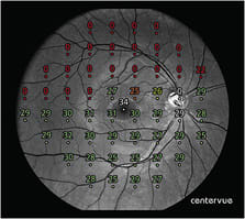
CENTERVUE
CenterVue COMPASS
Features:
- Combined VF and confocal retinal imaging
- Fundus-tracking technology reduces fixation loss.
- Full network capabilities
- No trial lenses are necessary due to auto-focus.
Link: bit.ly/CVCompass
ESSILOR
RETINA 800
Features:
- Fully automatic retinal imaging
- Fast and space-saving design, at 18 inches by 17 inches by 13 inches.
- Excellent image quality: clear and true color; 2.5 mm pupil size capability; 90° mosaic imaging
- Delegation-ready user interface: intuitive and direct connection with DICOM server
Link: bit.ly/Retina800

HEIDELBERG ENGINEERING
Spectralis OCT
Features: Spectralis is a modular diagnostic imaging platform that combines scanning laser fundus imaging with high-resolution OCT. It is easily upgraded to suit the needs of the practice. Key features include:
- Fundus imaging modalities: infrared, multicolor, bluepeak autofluorescence, blue reflectance (also known as red-free)
- Confocal optics provide a high-contrast, high-quality image, even in patients who have cataracts or other opacities.
- Patented TruTrack Dual Beam “Live” Eye Tracking tracks the eye to mitigate motion artifacts.
- Auto-rescan automatically positions follow-up exams in the same location to aid in detecting change as small as 1 μm.
- 10-layer visualization provides high-resolution OCT images from vitreous through choroid.
Link: bit.ly/Spectralis

KONAN
Nexy
Features: Robotic retinal imaging device, including:
- Fully automated hands-free operation
- Patented, polarized, high-fidelity imaging technology
- 45°, non-mydriatic camera that provides up to seven image locations with seamless mosaic
- Competitively priced
Link: konanmedical.com/nexy/

KOWA
Kowa nonmyd 8s Retinal Camera
Features: The non-mydriatic digital fundus camera offers:
- 3.3 mm minimum pupil diameter
- Multiple internal fixation targets
- High-resolution recordings of the fundus and anterior segment
- Upgradable DSLR cameras allow long-term use.
- Low-flash intensity for patient-friendliness
- The panorama function enlarges angle of view.
- Networking and connectivity for an efficient workflow
- Picture and documentation software VK-2 for data processing included.
- Software-guided stereo recordings assist with accurate glaucoma diagnosis.
Link: bit.ly/Nonmyd8s

NIDEK
MP3 and MP3S (Scotopic)
Features: MP3 and MP3S are automatic microperimeters with 12 MP high-resolution non-mydriatic fundus cameras, including:
- Wide measurement range
- Auto-tracking and auto-alignment
- Feedback exam for visual rehabilitation
- Fixation test
- Gray scale
- Easy image acquisition
- Scotopic microperimetry (factory-installed option)
Link: usa.nidek.com/microperimeter-mp-3/

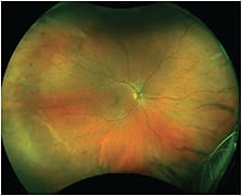
OPTOS
Monaco
Features: Monaco combines true single-capture, ultra-widefield (UWF) optomap images with an integrated 40° SD-OCT. Monaco produces a six-image overview showing color and autofluorescence optomap images and OCT scans of both eyes in as few as two minutes.
Monaco also features the OptosCloud, a secure, conven-ient, economical option for storing and reviewing images and diagnostic reports.
Optomap meets the definition of true UWF retinal images, as established by the International Widefield Imaging Study Group, by encompassing retinal anatomy anterior to the vortex ampullae in all four quadrants.
Link: optos.is/MonacoUWFandOCT
OPTOVUE
Vivicon Non-Mydriatic Ophthalmic Camera
Features:
- Easy-to-use with touch-screen tablet and automated image capture
- Photo montage, external image and manual modes
- Photo review, enhancement and export in choice of file formats
- Affordably priced for the everyday eye care practice
Link: optovue.com/products/vivicon

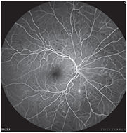
TOPCON
Topcon Maestro2
Features: The Topcon Maestro2 provides OCT and fundus imaging. The Maestro2, direct successor to the Maestro OCT, offers optometrists a user-friendly device with powerful imaging capabilities, including fully automated SD-OCT, true color fundus photography and the new Hood Report for Glaucoma. With one touch, optometrists can align, focus and capture the images they need, saving time and creating a better experience for patients and practitioners. The Maestro2 is also compact and space-efficient, offering significant imaging and reporting features with a relatively small footprint.
Link: https://www.topconmaestro.com/

VOLK
Pictor Plus
Features: The Pictor Plus is a portable, non-mydriatic, retinal camera that shoots high-resolution, detailed images in daylight or darkness. It is available in two modules: retina and anterior. The retina module allows the optometrist to examine and monitor conditions, such as glaucoma, diabetic retinopathy and AMD, through documentation of images, and facilitates patient education about disease status and the importance of regular eye exams. The anterior module helps with diagnoses by imaging corneal scratches, growths, rashes and other states present on the surface of the eye and surrounding areas. Additionally, the Pictor Workstation wirelessly transfers images from the device to a computer via Wi-Fi or USB, seamlessly integrating images into the EHR.
Link: bit.ly/PictorPlus

ZEISS
ZEISS CLARUS 700
Features: The Clarus 700:
- Captures ultra-widefield images in true color — that is, color closely approximating the natural coloration of the retina, as viewed through direct observation
- Provides quality images
- Offers a complete suite of modalities, including fluorescein angiography
- Also possesses advanced features, such as modalities of true color with RGB separation and stereo image pairs.
Link: bit.ly/CLARUS
Resources
IN ADDITION TO THE COMPANIES LISTED, CONSIDER CONTACTING PROFESSIONAL ORGANIZATIONS, SUCH AS THOSE BELOW, FOR INFORMATION ON IMAGING AND DIAGNOSTIC DEVICES.
→ OPTOMETRIC GLAUCOMA SOCIETY: The society’s objectives include “to promote education of optometrists related to all aspects of glaucaoma,” the “About Us” page says. Find more information on the web at optometricglaucomasociety.org .
→ OPHTHALMIC PHOTOGRAPHER’S SOCIETY: This organization provides “primary and continuing education in the field of ophthalmic photography. . .” among other main objectives. More information about it can be found at opsweb.org .
→ OPTOMETRIC RETINA SOCIETY: The retina society’s website says, “We provide updates in retinal disease that are important to primary care optometrists.” Find out more about those updates and resources at optometricretinasociety.org .