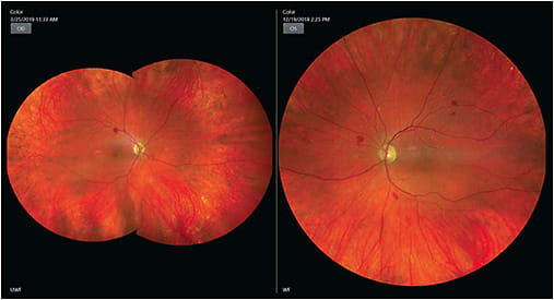
This SLO image shows severe non-proliferative retinopathy with multiple microaneurysms and intraretinal hemorrhages, cotton-wool spots, and exudates. Image taken from Heidelberg Engineering SPECTRALIS OCT.
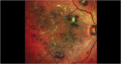
This image was taken from a 57-year-old Black male with severe, non-proliferative DR. The patient has had diabetes for 17 years, and takes insulin and metformin. Image taken with the iCare DRSplus.

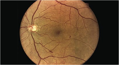
This patient has moderate diabetic retinopathy, dot and blot hemorrhages, and an occasional hard exudate. Image taken with the Kowa Nonmyd WX3D.
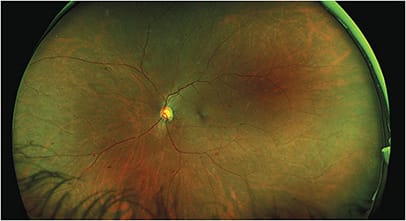
An ultra-widefield optomap image of a patient who has proliferative diabetic retinopathy; despite the overall quiet appearance, a closer look shows neovascularization of the disc and intraretinal hemorrhages. Image taken with the Optos California.
A color fundus image, taken with the Topcon Triton SS OCT Color Fundus Camera, showing scattered intraretinal dot and blot hemorrhages, isolated cotton wool spot superior temporal, venous caliber changes, and intraretinal lipid centrally.
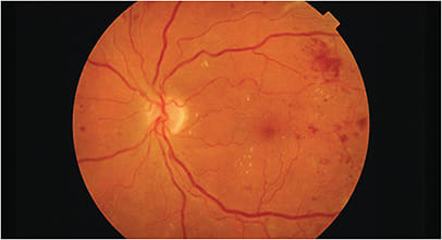
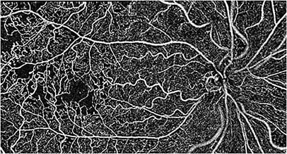
A montage image taken of a 54-year-old diabetic Black female who has reduced vision and a BCVA of 20/40 OD. The image was captured using Visionix’s Optovue Avanti, with the AngioVue software.
An image of scattered dot and blot hemorrhages and microaneurysms. Image taken using the Zeiss Clarus 500 fundus imaging system.
