Because retinal diseases, such as diabetic retinal disease (DRD), age-related macular degeneration (AMD), vascular occlusions, hypertensive retinopathy, vitreoretinal interface abnormalities (e.g., vitreomacular traction syndrome, epiretinal membranes), pathologic myopia, and macular hole are significant causes of visual impairment and blindness, it’s essential we educate our patients about these conditions. After all, an educated patient is the more likely to take an active role in the management of their disease — a requirement to increase the likelihood of staving off vision loss.
That said, with the time-intensive requirements of managed care, it can be challenging to personally provide the education required to set patients up for success. With that in mind, here are tips on how to provide this needed education efficiently.
USE STAFF
Whether it’s the tech who delivered the patient to your chair, or a trusted scribe, these staff members can step in and provide education to patients on the diagnosed retinal disease.
Obviously, a trained and knowledgeable staff is essential to accomplishing this. This training can occur from staff meetings, lunch and learns, attending education offered at trade shows, webinars, podcasts, meetings with pharmaceutical companies, and shadowing one or more doctors for a time.
For example, if you have prescribed a nutritional supplement for a dry AMD patient, a tech or scribe can explain to this patient the role the supplement will play in enabling them to maintain their current vision. Depending on the doctor’s choice of ocular nutritional supplement, the staff may briefly educate the patient on the supplement’s benefits. For example, my staff discuss the findings of the two Age-Related Eye Disease (AREDS) studies and how a recently published 10-year follow-up study affirms that AREDS 2 with lutein remains an effective and safe choice.1 Additionally, staff should discuss the importance of patient compliance to your specific prescription.
Another example: If the patient has a history of diabetes, but is not showing signs of diabetic eye disease, a staff member can explain the “ABCs” of diabetes: (HbA1c ≤7), (B) blood pressure ≤ 130/80 mm Hg, and (C) cholesterol, and (s) (smoking cessation) to prevent diabetic eye disease onset.
One more example: For those patients who have severe retinal disease/vision loss, staff can go into greater detail on the appropriate treatment options (e.g., intravitreal injections, laser, or surgeries) and, if appropriate, low vision services, as well as calm fears by discussing their related procedures for use.
Pro tip: Before leaving the exam room, I suggest you introduce the staff member who will be providing the education to the patient. The reason: Doing so sends the message to the patient that you, as the expert, trust the staff member to deliver the information the patient needs to stay on top of their diagnosis.

SHOW IMAGING/DIAGNOSTIC TESTING
Verbally explaining a patient’s retinal condition is far more time consuming and confusing for the patient than using the findings of a diagnostic test or a clinical image to provide patient education.
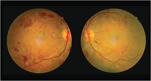
Fundus photography not only aids in the diagnosis and management of retinal disease, it is also an excellent tool for efficient patient education, as it illustrates pathology. As many retinal diseases involve the peripheral retina, ultra-widefield imaging is also an excellent tool to capture most of the retina in a single image, another time saver. (For a list of retinal disease diagnostic devices, see bit.ly/OMRetinaDiagnostics .)
A script to follow is “Diabetes may cause bleeding and blood vessel leakage — as well as other changes — in your retina. This device takes pictures of your retina so that we can detect such changes.”
As a brief yet related aside, showing patients imaging and diagnostic test results emphasizes to them the importance of adhering to preventative measures (good glycemic and blood pressure control, diet, and nutrition) to delay or prevent vision-threatening complications, Additionally, these technologies stress the importance of regular dilated eye exams in detecting changes before visual loss manifests.
PROVIDE PATIENT EDUCATION MATERIALS
Another method of providing patient education on retinal disease efficiently is to send patients home with or email them related informational materials. These can include large-print FAQ-type sheets (see bit.ly/OMAMDFAQ and bit.ly/OMDRNEI ), as examples); specific treatment instructions (utilizing step-by-step graphics); and videos (see bit.ly/OMDRVideo and bit.ly/OMAMDVideo , as examples).
A bonus: As patients remember roughly half of what they hear during their visit with their doctor, according to a recent Brown University School of Public Health study,2 these related informational materials can fill in the blanks for patients, re-emphasizing to them the need for following the discussed management and follow-up schedule.
Retinal Disease at a Glance
DRD. Diabetes retinal disease (DRD) — diabetic retinopathy (DR) and diabetic macular edema (DME), are the leading causes of visual impairment in individuals of working age.3
AMD. It is estimated that between 11 to 15 million people have some form of AMD in the United States, and that number is expected to increase to 22 million by 2050.4-5
DELIVERING WHAT IS NEEDED
It’s a given that patient education is essential in the management of retinal disease. A better-informed patient is the more likely to take an active role in preventing and reducing the vision loss from the diagnosed retinal disease. While time is at a premium in practice, I have found that the above courses of action work to deliver patient education efficiently, while fostering the importance of their participation in their care. OM
References
1. Emily Y Chew; Traci E Clemons; Tiarnan D L Keenan; Elvira Agron; Claire E Malley; Amitha Domalpally. The Results of the 10 Year Follow-on Study of the Age-Related Eye Disease Study 2 (AREDS2). ARVO Annual Meeting Abstract. 2021.Volume 62, Issue 8
2. Brown University. School of Public Health. Do You Remember What Your Doctor Said? It May Depend on Several Factors. https://www.brown.edu/academics/public-health/news/2018/08/do-you-remember-what-your-doctor-said-it-may-depend-several-factors. Accessed November 2, 2022.
3. Centers for Disease Control and Prevention. National Diabetes Statistics Report website. https://www.cdc.gov/diabetes/data/statistics-report/index.html. (Accessed October 30, 2022.)
4. Wong WL, Su X, Li X, Cheung CMG, Klein R, Cheng CY, et al. Global prevalence of age-related macular degeneration and disease burden projection for 2020 and 2040: a systematic review and meta-analysis. Lancet Glob Health. 2014. 2: e106–e116.
5. Emily Y Chew; Traci E Clemons; Tiarnan D L Keenan; Elvira Agron; Claire E Malley; Amitha Domalpally. The Results of the 10 Year Follow-on Study of the Age-Related Eye Disease Study 2 (AREDS2). ARVO Annual Meeting Abstract. 2021.Volume 62, Issue 8
MORE ON RETINAL DISEASE


ARE ANY APPS AVAILABLE TO HELP PATIENTS WHO HAVE VISION LOSS?

HOW CAN ODs DIAGNOSE AMD EARLY?

HOW CAN ODs IDENTIFY THE RETINAL MANIFESTATIONS OF SYSTEMIC MEDICATIONS?


