Note: This article is one in a continuing series on diagnostic devices
When I first began practicing 25 years ago, there were no OCTs, widefield retinal cameras, handheld ERGs, or similar items in my clinic. Today, though, the tools used to improve the patient experience seem to multiply every year.
We have undoubtedly entered a new era in optometry; an era marked by the advent of ever-more-sophisticated diagnostic and treatment devices.
In this article, I’ll show you how patient care at my practice has been improved by some of these technologies. The devices I will discuss here are largely used in my diagnosis, management and treatment decisions for “back of the eye” conditions. I have found that they have been invaluable in allowing me to make better clinical decision. They have increased the efficiency of our practice and are appreciated by our patients for their ease of use and educational value.
BENEFIT TO PATIENTS
Easier exams. Obviously, any device that makes the exam easier can help make the patient more comfortable and make the practice run more smoothly. One recently purchased device that helps my practice with this is a handheld electroretinogram (ERG). This device (unlike older ERG systems) is small, handheld, and technician-driven. Patients love that the test does not require a “clicker” and can be done in 15 seconds to 90 seconds per eye; another advantage of being “clicker-less” is that the handheld ERG can take an objective measurement of the patient eye, without any response from the patient which would increase the chance of human error. The device is able to assist with evaluating the functioning of the retina in patients who have diabetic retinopathy, possible glaucoma, and other congenital retinal and optic nerve diseases.
I have begun using the ERG for many patients who have diabetic eye disease and for folks who struggle with more traditional subjective testing that often yields unreliable results (threshold visual field in certain glaucoma patients). With the advent of a handheld device with simple lower lid electrodes, I can get very reliable and repeatable objective functional data on patients who have a wide range of retinal diseases, such as diabetic retinopathy, congenital retinal diseases, glaucomatous optic atrophy, and age-related macular degeneration (AMD).
Earlier diagnosis. Any device that can allow for easier, quicker diagnosis gives a better chance for preserving sight. This was a major benefit of adopting dark adaptation testing, which allowed my practice to detect AMD often prior to clinical signs of the disease. Adding dark adaptation to our practice and using the device as a screening tool in those who have a night vision complaint has allowed us to find significant macular disease, often in the absence of obvious structural damage.
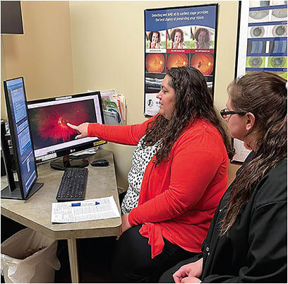
Patient education. Pictures can be incredibly helpful when educating a patient on their condition. In my experience, the level of interest and the ownership of the patient in the management plan goes up considerably when they can visualize the results of their efforts and see the progress of their health in a quantifiable way.
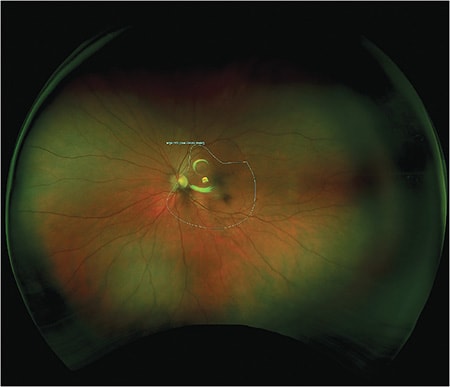
One device that reinforced this for me was the widefield fundus camera. When I began using it in 2004, it was the first time I was able to visualize retinal disease and show my patients the status of their eye health in beautiful living color! There was a significant difference between describing diabetic retinopathy or a large cup-to-disc ratio to a patient vs. showing them an image of their retinal findings. Diabetic patients could see their retinopathy improving after initiating better lifestyle choices and a better diet, and patients could clearly see what we referred to when we used terms like “the Weiss’ ring” or “posterior vitreous detachment.”
Patients loved the fact that images could be taken through an undilated pupil, and, for visits where the image was not covered by Medicare, most were more than happy to spend additional money to “see” the back of their eye and stay updated on their retina health. The patient was happy, I was thrilled, and the camera paid for itself in very little time. To this day, many patients will prefer to spend some extra money to have an image taken even when we insist that they must also be dilated.
No need to visit a second practice. Patients, like all of us, are busy, and appreciate it when we can save them a trip to a separate medical office. This is one of the benefits we found in adding optical coherence tomography (OCT). The OCT allowed us to image the layers of the retina in cross section – this not only opened a new world of diagnostic awesomeness, but allowed us to confidently identify areas of the retina affected by disease, thus allowing for more timely and appropriate referrals to our retinal specialist friends. In many cases, our patients could be monitored and managed in our office; this saved patients the inconvenience of making a separate visit to a retina specialist, which thrilled our patients.


Allowing Patients to be Proactive. Many of our patients with AMD have family or friends who have lost vision to the condition. They want to find a way to be proactive with their condition, and that’s where we’ve found home testing for AMD helpful, as it can notify the practice when a patient may have a dry-to-wet conversion. The cost of the devices and the monitoring service are covered by Medicare. Our AMD patients are usually extremely grateful to have this chance to take better control of their condition.
BENEFITS FOR THE PRACTICE
Keep patients in your clinic. Another advantage of introducing OCT at our practice is that it allowed us to retain the patient and collect the fees associated with the testing and the office visit. When we did refer out to another clinic, our retinal doctors came to appreciate that the referrals they received from our clinic were necessary ones with a treatable condition.
More confidence in testing. Having technology that can very accurately diagnose patients, as is the case with dark adaptation and AMD, lets us be more certain we’re calling the patient’s diagnosis correctly the first time. This gives peace of mind that we are likely initially making the correct diagnosis for our patients.
Quick response to AMD conversion. Adding home monitoring technology allowed our office to act quickly when an AMD conversion is likely, significantly improving visual outcomes for patients. Our office is also able to capture the revenue from the office visits.
A WIN-WIN
The use of modern technology has surely made my practice a destination for patients worried about their retinal health. I have had numerous patients refer family and friends to our practice simply because we have some of the most modern technology, allowing us to not just improve their treatment but their whole patient experience.
It is surely a win-win for our patients and our practice as we continue to grow the medical model. OM


