This article was originally published in a sponsored newsletter.
In degenerative diseases, the function or structure of affected tissues or organs changes for the worse over time.1 In addition to elucidating the nature of macular degenerative lesions, early AMD studies recognized several clinical findings located beyond the macula, into the retinal periphery.2,3 However, for the past several decades, these peripheral associations were ignored as researchers zeroed in on treating the macula with therapies ranging from laser to photodynamic therapy to nutrition and anti-VEGF therapy.
In 1997, technology was created that allowed a 130° view of the retina. It consisted of a wide-angled camera, a contact lens and multiple lens attachments.4 In 2000, Optos introduced widefield fundus imaging technology, which provided a 200° view of the retina.4 Other companies followed this lead, and now there are several widefield and ultra-widefield (UWF) devices available to clinicians.
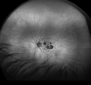
Fundus autofluorescence (FAF) was first used for in vivo fundus imaging in 1995 when Delori et al implemented a spectrophotometer to characterize the intrinsic autofluorescent properties of the human retinal pigment epithelium (RPE).5 Over time, there have been many technological advancements that have allowed for the development of numerous commercially available FAF devices, including widefield FAF (see Figure 1). The use of widefield fluorescein angiography has enabled easier identification of non-central neovascular AMD (nAMD) lesions, and OCT-angiography is trending in a more widefield direction as well.
These recent advances in fundus imaging have sparked a renewed interest in peripheral retinal degenerative disease. Emerging evidence shows that clinical features associated with AMD are not exclusive to the macula. In a meta-analysis of 12 studies that included 3,261 eyes, Forshaw and colleagues studied peripheral retinal lesions in eyes with AMD.6 The most observed peripheral lesions were drusen, atrophy and changes to the RPE. In eyes with AMD, peripheral lesions were found in 82.7% of eyes (confidence interval, 78.4% to 86.7%) compared with 33.3% of healthy eyes (confidence interval, 28.3% to 38.5%).6
Lewis found that reticular degeneration of the RPE (RDRPE) and AMD were concomitant manifestations of the aging process, and RDRPE may be a significant risk factor associated with AMD.2 Evaluation of the peripheral fundus is of clinical value when assessing patients with macular degenerative abnormalities.

Key Clinical Takeaways
- Although macular changes are the hallmark of AMD, there has been renewed interest in peripheral findings in the last decade.
- Peripheral changes have a higher prevalence in AMD patients compared to the general population. This finding has led to an increased focus in exploring their significance.
- UWF imaging, with and without FAF, has enabled a better understanding of peripheral changes in AMD.
- The presence of peripheral lesions may contribute to impaired visual function in these patients.
Reference(s):
- National Cancer Institute. Degenerative disease. Accessed February 24, 2024. https://www.cancer.gov/publications/dictionaries/cancer-terms/def/degenerative-disease
- Lewis H, Straatsma BR, Foos RY, Lightfoot DO. Reticular degeneration of the pigment epithelium. Ophthalmology. 1985 Nov;92(11):1485-1495. doi:10.1016/s0161-6420(85)33829-0
- Lewis H, Straatsma BR, Foos RY. Chorioretinal juncture. Multiple extramacular drusen. Ophthalmology. 1986 Aug;93(8):1098-1112. doi:10.1016/s0161-6420(86)33615-7
- OPTOS. OPTOS home. Accessed February 24, 2024. www.optos.com
- Delori FC, Dorey CK, Staurenghi G, Arend O, Goger DG, Weiter JJ. In vivo fluorescence of the ocular fundus exhibits retinal pigment epithelium lipofuscin characteristics. Invest Ophthalmol Vis Sci. 1995 Mar;36(3):718-729.
- Forshaw TRJ, Minör Å, Subhi Y, Sørensen TL. Peripheral retinal lesions in eyes with age-related macular degeneration using ultra-widefield imaging: a systematic review with meta-analyses. Ophthalmol Retina. 2019 Sep;3(9):734-743. doi:10.1016/j.oret.2019.04.014


