This article was originally published in a sponsored newsletter.
For the most part in the setting in which I practice, my day is spent seeing sick eyes. These eyes may have chronic conditions such as glaucoma or macular degeneration, but they may also have acute conditions that need more immediate care. In both chronic and acute conditions, OCT technology has given us the ability to quantify the amount of change seen over time. In some cases such as glaucoma, this change equates to worsening. However, it can also equate to improvement in other conditions. Here is one such case of improvement over time, and the big picture perspective that the Spectralis provides.
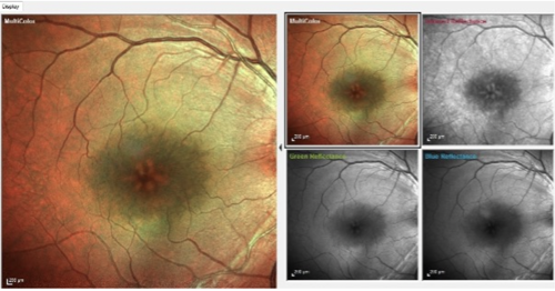
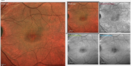
This 77-year-old African American patient recently underwent cataract surgery in both eyes. The initial post-op visit at one week demonstrated significant improvement in vision in both eyes, but at his three-month visit, he complained that his vision seemed to have gotten worse. A quick look at the anterior chambers demonstrated mild cell and flare bilaterally, and the posterior pole evaluation demonstrated the classic petalloid appearance associated with cystoid macular edema (CME; see Figures 1 and 2).
Before OCTs became available, we had to depend on detailed fundus views in vivo to make diagnoses in practice. Subtle macular edema was (and still is) difficult but not impossible to see without OCT. Significant macular edema was obvious on fundoscopy; though, not surprisingly, visual acuity was greatly reduced in cases of significant edema, such as this one.
It can be good to have a record of the fundus appearance aside from OCT, which we can get with the Spectralis MultiColor imaging system. Using three wavelengths of light, the MultiColor image system highlights different layers of the retina, from anterior to middle to the choroidal layer. When these three images are stacked together, we have the false color images, seen here on the left side.


Aside from the macular edema, note the nerve fiber layer wedge defects in both eyes, especially on the blue reflectance image. They are consistent with this patient’s glaucomatous damage.
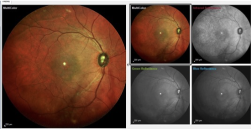
The MultiColor images give us a global picture of the posterior pole. Figures 1 and 2 are 30° images, but these images can also be obtained in 55° views (see Figure 3), which are helpful in providing an even broader record of the fundus, especially in diabetic retinopathy cases.

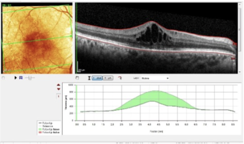
Management of post-operative CME is straightforward, and our OCT images help us to clinically determine improvement, and ultimately, confirm resolution of the condition. For instance, Figure 4 shows the OCT images of this patient’s right eye mid-way through the treatment plan, with significant reduction in the macular edema from baseline. While there is still a significant amount of edema remaining, we can now measure improvement over time at the micron level.

The Spectralis proves itself to be a versatile clinical tool with both the big picture and the micron-level picture. These images are invaluable in managing posterior segment conditions that we can get in the same sitting for patients.


