This article was originally published in a sponsored newsletter.
Soft contact lenses have become more streamlined for practitioners to fit and prescribe, allowing most patients to be fit relatively successfully with reproducible single vision, toric and multifocal designs. The companies that produce contact lens technologies frequently offer parameters, such as the power, base curve and diameter, for lens fitting. However, the depth of the bowl (i.e., the sagittal depth) is an often overlooked factor that can influence both comfort with and vision through the lenses.
Take, for example, a patient who has a shallow sagittal depth but is fit with a soft lens that has a significantly greater sagittal depth than what is required for their cornea. This patient may experience poor visual quality because of the soft lens vaulting over the surface of the eye, as well as visual fluctuations with blinking. Immediately after they blink, they may have better vision, but as they keep their eyes open between blinks, their visual quality reduces again. Fortunately, advances in optical coherence tomography (OCT) provide us with the ability to measure sagittal depth at different chords that we can determine as clinicians. We can also refer to Pacific University’s resource that has measured sagittal depth for most contemporary soft contact lenses.1 This case demonstrates how OCT technology can be utilized to best match the appropriate sagittal depth with each patient.
Case in Point
A 30-year-old female came in to see me for an eye exam. Her chief complaint was that she had poor vision out of her current contact lenses. She said she hadn’t been able to see well out of them since she was refit with them two months prior.
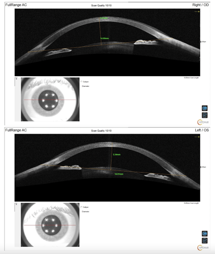
At the slit lamp, the lenses seemed to fit. We attempted over-refraction over the contact lenses and the best corrected vision acquired was OD 20/30 and OS 20/30. Knowing the lenses that the patient was wearing, we looked up the sagittal depth on the Pacific University sagittal depth chart and found that it was 3,688 μm.1 We measured an 18 mm anterior segment horizontal cross section OCT on the right and left eyes using the Optovue Solix by Visionix (Figure 1), and drew a chord of 14.0 mm (the diameter of the lens that the patient was wearing).
The sagittal depth from the 14.0 mm chord to the anterior surface of the cornea was 3,410 μm for the right eye and 3,360 μm for the left eye. At that point, it was obvious that the sagittal depth of the patient’s current lenses was too great for her eyes. We placed a lens with a sagittal depth of 3,434 μm and appropriate power, and her vision immediately improved.
Understanding the importance of sagittal depth and its influence on soft contact lens vision is critical. Fortunately, advanced OCT technologies like the Optovue Solix provide us with opportunities to measure sagittal depth and provide our patients with better contact lens outcomes.

Reference(s):
1. van der Worp E. Pacific University SCL sagittal depth story. Pacific University Oregon Research Repository. January 2021. Accessed May 31, 2024. https://commons.pacificu.edu/work/ns/380b99ad-5f0b-4e77-a286-a8d1f74bdfe9


