In the world of OCT interpretations, we often concentrate on a specific area of the retina or optic nerve, looking at a particular pathology that may only be a few microns in size. The resolution of our OCT instruments is quite impressive and OCT technology has an invaluable place in our clinics across a spectrum of diseases.
Sometimes, though, it’s good to step back from the micron level and look at the whole picture. Of course, there are OCT instruments such as the Spectralis (Heidelberg Engineering) that can take widefield OCT images of the posterior pole to give us a more global perspective of a particular B-scan image. But there is also the need for widefield imaging of the posterior pole to help us to put things into perspective. Here is an example of such a case.
This pleasant 72-year-old female has been a patient of mine for the past ten years, initially coming to me to establish care after moving to the area. Lifelong T2 diabetes is significant in her history, along with multiple laser procedures to both eyes secondary to proliferative diabetic retinopathy. While her diabetes has been stable for the past 20 or so years, the damage done earlier in her life has resulted in significantly compromised vision OU. Over the years, she has adapted to the low vision situation, but certainly wishes things were different. The good news is that she hasn’t worsened, but the bad news is that she won’t get better.
Visual acuities are FC at 3 feet OD and 20/800 OD. OCTs of the posterior segment are part of her 6-month follow-up visits to look for any subtle changes that may occur and compromise vision further, especially in the OS. A dilated fundus exam is also part of each visit. When I saw her last week, her entire fundus reminded me to step back and see the big picture.
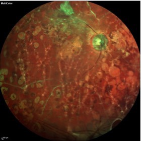
Utilitarian instruments such as the Spectralis OCT offer several ancillary imaging techniques—such as fundus autofluorescence and, in this case, multicolor imaging—that are managed from the same hardware platform. Multicolor imaging is a form of multimodal imaging, whereby the Spectralis unit images the posterior pole using three wavelengths of light to highlight the deep, middle and superficial layers of the retina. These images can also be taken in a 55゚wide format, thereby giving us the “big picture.”
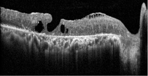
Note the OCT details of the macula in the right eye, in particular the significant disruption of the neurosensory retina and the underlying retinal pigment epithelium (RPE), in Figure 1 below.

In Figures 2 and 3 below, we see a widefield multicolor image of both posterior poles, which demonstrates the extent of the damage in both posterior poles. Therefore, we have a clearer perspective of the basis for her severely reduced vision.


In Figure 2, note the off-centered posterior pole image, owing to the patient’s inability to fixate in this eye. And with better acuity in the left eye, we have a well centered image in Figure 3 that gives us the “big picture.”
Micron resolution is fantastic in the Spectralis, but sometimes we need to step back to get a broader perspective, and the Spectralis can assist nicely with this endeavor as well.


