This article was originally published in a sponsored newsletter.
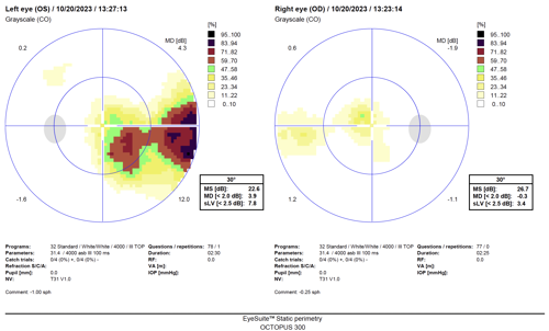
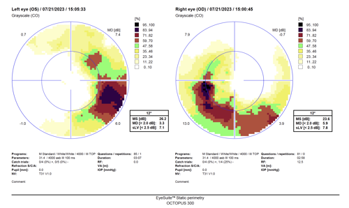
The presenting patient is a 61-year-old white female with severe normal tension glaucoma in both eyes. The 30-2 Octopus Visual fields below (Figure 1) show a shallow paracentral scotoma in the right eye and nasal step in the left eye. The field depression is near 10° within fixation on the left eye and a dense, larger paracentral scotoma is within 5° in the right eye on the macular visual field analysis.
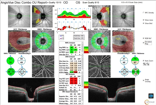
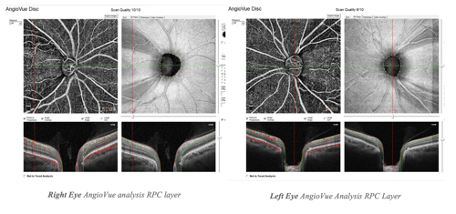
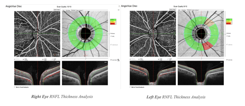
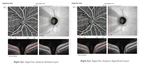
Evaluating the optic nerve with the Optovue Solix Disc Combo Report 9 (Figure 2), the right eye has green disease of the retinal nerve fiber layer (RNFL) due to normal appearance of NFL on statistical analysis. However, the ganglion cell complex (GCC) thickness report (Figure 3) shows the extended view of NFL thinning extending from the optic nerve, which is consistent with the macular visual field loss. Evaluating the AngioVue Disc report of the right eye, the thinning of the vessel density correlating to the NFL wedge defects inferior and superior temporally can be appreciated especially at the radial peripapillary capillary (RPC) and superficial layers. Examining the en face AngioVue Disc also shows the correlating NFL wedge defects. The left eye has a similar appearance, but does not have green disease and displays the thinning of the NFL thickness that correlates with the field loss.
Using the Optovue Solix AngioVue report and evaluating the vessel density loss, as well as the en face and extended GCC analysis, can help rule out green disease, especially when the patient has severe vision loss that may be missed with conventional OCT analysis reports and 30-2 visual field testing. This information is essential when determining the stage of glaucoma and how aggressive treatment needs to be to prevent further central vision loss.
