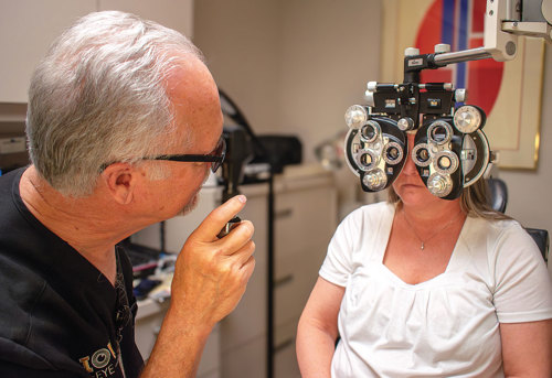
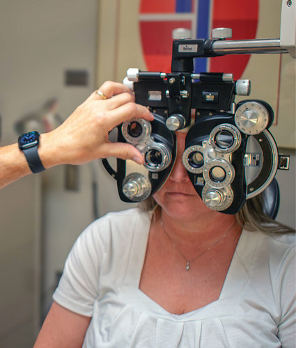
As optometry moves more toward ocular disease management and treatment, we must not forget the value of a careful refraction. What follows is a review of the steps needed by you or your refractive staff to achieve an accurate refraction every time.
First things first
This starts with performing refractions with the exam room lights on. The reason: Doing so precludes overminusing your patients, which often happens in a dark exam room due to low light-induced refractive shifts (night myopia).
Something else to keep in mind: If the exam room is less than optical infinity, which is 20 feet, vergence and accommodation will apply. To calculate vergence, use the formula 1/x (m), or 100/x (cm) or 40/x (in). In a 10-foot exam lane, the vergence demand is 40/120 = 0.33 D. Thus, if testing is occurring in a 10-foot exam lane, the patient is getting an additional -0.33 D of refracting power in this lane. To counteract this, you should apply additional minus power to the prescription that was found in the phoropter to move the patient’s focal point to optical infinity. For example, you should add -0.25 D for a 10-foot exam room. Add -0.50 D for a six-foot exam room.
Additionally, you or your team will want to obtain an objective assessment of the patient’s refraction via autorefraction or retinoscopy, prior to starting the subjective refraction. (A caveat: Those who are physically unable to sit up, such as babies, and patients who have nystagmus or reduced central vision, do not make good candidates for autorefraction. When seeing such patients, you will need to use a retinoscope and perform a trial frame refraction to accurately determine their refractive needs.) I caution against using the patient’s previous prescription to obtain the objective refraction. The reason: It doesn’t provide any objective information about the patient’s presenting refractive error. (Remember: Vision can change regardless of the prescription in their current pair of glasses.) (See “Performing the pinhole acuity test,” below.)

Compensating for astigmatism
To determine the patient’s cylindrical correction, you or your team can employ the Jackson cross-cylinder (JCC) technique. How to perform it:
If autorefraction or retinoscopy reveals 1.00 D or more of cylinder, or the patient has a mostly cylindrical refractive error, you or your team want to check the cylinder axis first. The reason: It’s not possible to determine the patient’s correct cylindrical power when the axis is incorrect.
To find the patient’s true cylinder axis, you or your team line up the JCC flip knobs with the cylinder axis, sometimes marked as an “A” on the phoropter, so that the JCC’s white and red dots straddle the cylindrical axis by 45° on either side.
Performing the pinhole visual acuity test
For individuals who do not have any type of ocular disease, a pinhole aperture can be useful in determining a refractive error or a required refractive change. A pinhole diameter of 1.2 mm is ideal for clin-
ical purposes, as it is effective for
+/- 5.00 D refractive errors.
A pinhole improves VA by reducing blur circle size on the retina. The outcome is an improvement in VA. Macular dis-ease patients and patients who have other central vision-affecting conditions may have similar or even decreased VA when using a pinhole. The reason: The reduced amount of light entering through the pinhole decreases the clarity of the chart.
Also, using eccentric fixation through a pinhole can be difficult. Thus, individuals who have ocular disease should not be told that a spectacle correction change will not improve their vision, based solely on them looking through a pinhole. Careful retinoscopy and a trial frame refraction is required to find out whether a patient who has pathology-induced vision loss will find value from a spectacle correction change.
Next, you or your team instruct the patient to look at one-to-two lines above their best VA (determined during the baseline maximum plus to maximum VA), flip the axis between the red and the white dots, and tell the patient “I am going to give you two choices. Tell me which lens choice is clearer, in understanding that neither choice will be crystal clear.”
For patients who have 2.00 D of cylinder or less, you or your team rotate the axis closer to the red dot (for minus cylinder phoropters), starting with 15° increments. This is known as “chasing the red” because the red dot places the axis closer to the patient’s true cylinder axis, making the letters clearer for the patient. For patients who have greater than 2.00 D of cylinder, you or your team employ 5° increments, reducing the increment size after a reversal to 5-3-1° until you have refined their cylinder axis.

It’s time to check the cylinder power. To accomplish this, you or your team manipulate the JCC to make its white or red dots correspond with the patient’s cylinder axis, and ask the patient “Is lens choice one or lens choice two clearer?”
If the patient picks the white dot, you or your team remove -0.50 D of cylindrical power and remember to add -0.25 D of spherical power to keep the spherical equivalent. If the patient opts for the red dot, apply -0.50 D of cylindrical power, and add +0.25 D of spherical power to keep the spherical equivalent.
Should the patient pick the red dot after the white one or vice-versa, known as reversal, you or your team change the cylinder power by 0.25 D in the opposite direction of your prior change. (You are not required to change the spherical power for the 0.25 D adjustment.)
Now, recheck the cylindrical power to determine whether the patient needs more or less of it. The goal is the least amount of cylinder power that provides the best vision. Once you or your team have refined the cylindrical power and axis, withdraw the JCC, and instruct the patient to read the smallest line of letters they are able to.
Of note: There are two scenarios where you won’t perform the axis check first: (1) When retinoscopy or autorefraction reveal the patient has no astigmatism and (2) when the retinoscopy or autorefraction reveals 0.75 D of astigmatism or less. To be sure these findings are correct, you want to check cylindrical power first to see whether the presence of astigmatism is legitimate. If no astigmatic correction was found by autorefraction or retinoscopy, you or your team can check for cylindrical power, by orienting your JCC for power at 90° and 180°, and ask the patient “which is better: one or two?” If the patient is indifferent, use 45° and 135°. If the patient picks one, apply -0.50 D cylinder at the preferred axis (red dot axis), and +0.25 D sphere to keep the spherical equivalent.
Now, refine the cylinder power and axis employing the described standard JCC technique. Record the patient’s best VA. Finally, cease occluding the left eye while simultaneously occluding the right eye. Do the same for the left eye and start by employing the patient’s initial maximum plus to maximum visual (MPMVA) acuity. (See “Achieving initial MPMVA,” below.)
Achieving initial MPMVA
To ensure you haven’t overminused the patient during retinoscopy or autorefraction, it’s important to obtain their baseline maximum plus to maximum VA (MPMVA).
To accomplish this, start by occluding the patient’s left eye. Next, show several letter lines in the ranges of 20/20 to 20/50 or 20/15 to 20/40 on the Snellen chart, and instruct the patient to read to you the smallest line of letters they are able.
Expecting the patient can read the presented letters, apply +0.75 D to the phoropter. The outcome of this action should be two-to-three lines
of vision loss.
Now, ask the patient whether they’ve experienced any vision loss. If they reply “no,” apply an additional +0.75 D to the phoropter, and ask again, as you want to make sure the patient has experienced a two-to-three-line reduction in vision from where they started.
Next, deliberately reduce (less plus more minus) the power in the phoropter, in 0.25 D steps until the patient says they can see the 20/20 or 20/15 lines, or they say they’ve experienced no better vision. For every -0.25 D added in the phoropter, the patient should achieve one line of improvement on the Snellen chart.
Finding the right balance
You or your team should work to obtain binocular balance when the patient’s VA is relatively the same in each eye. In addition to making sure the refractive power is balanced between the two eyes, another benefit of binocular testing is that it will allow you to determine whether the patient has diplopia, which can develop with age as binocular fusion abilities decline.
To achieve binocular balance, you can use the Risley prism on the phoropter or the fogging and alternate occlusion technique.
Regardless of whether you use the Risley prism or fogging and alternate occlusion, neutralize eye dominance by adding +0.75 D sphere to both eyes to blur the patient’s VA to the levels of 20/30 to 20/40.

To use the Risley prism in the phoropter, add three prism diopters base up to the front of the right eye, and add three prism diopters base down to the front of the left eye. The outcome: The lower image will be seen through the right eye, and the upper image will be seen through the left eye. Instruct the patient to disregard any brightness differences they may note, and to tell you which image looks clearer. Apply +0.25 D to the clearer eye to fog it more. Now, instruct the patient to tell you which image is clearer, and apply +0.25 D to the eye that sees the clearer image.
When both sets of letters look the same to the patient, or the patient’s dominant eye sees slightly clearer than their non-dominant eye, you’ve achieved binocular balance.
To use the alternate occlusion technique, after fogging both eyes, as noted above with +0.75 D, alternately cover one eye and then the other eye while instructing the patient to tell you which eye sees the Snellen chart more clearly. Then, apply +0.25 D to the clearer eye to fog it more. When both sets of letters look the same to the patient, or the patient’s dominant eye is slightly clearer than their non-dominant eye, you’ve accomplished binocular balance.
Once you or your team have finished the binocular balance, apply -0.25 D OU one step at a time to return the patient to their best VA. If the patient doesn’t experience improvement in VA, do not provide additional minus spherical power. (See “Refracting with the red-green test,” below.)
Refracting with the red-green test
Typically, I use this test only after the binocular balance to ensure that I haven’t overminused the final prescription.
With this test, if the letters on the green side of the chart appear blacker, you add +0.25 D. If the letters on the red side appear blacker, you add -0.25 D.
When the letters appear equally black on both the red and green side, you’ve reached the endpoint.
Correcting near vision
Although this tends to be on our radar in patients older than age 40, correcting near vision should always be based on a patient’s near vision complaint and not their age.
To correct for near vision, you or your team should use the plus build-up method to determine the add power needed. As the add power increases with age, it is important to remember that stronger is not always better because higher add powers (over +2.50 D) require a closer working distance than many people can tolerate.
The use of computers, particularly with multiple monitors, as well as smart phones and tablets have resulted in the need for many individuals to require more than one prescription for their different visual demands. The days when people could comfortably function with a two or three focus lens (bifocal or trifocal) have passed for the majority of our presbyopic population. Even a progressive addition may prove inadequate for someone working on two or three monitors. In this case, a single vision computer prescription focused for the longer working distance of the computer monitor(s) can greatly add to the user’s visual comfort. Additionally, the standard 16-inch reading distance of the past is no longer the preferred focal distance when looking at smart phones or tablets, as well as for playing music. Progressive lenses, as well as single-focus lenses designed for the specific viewing distance for these tasks are often the solution for those experiencing visual asthenopia with their standard multifocal prescription.
Requirements for providing the prescription
Consider the following before prescribing a new prescription:
• Your patient should be seeing better than with their current prescription or entrance VAs.
• The amount of change in the sphere, cylinder, and axis is consistent with the improvement in VA found for each eye.
• The add power should be consistent with what you would expect for the improvement in vision in the patient.
Subjective refraction is extremely important and valuable to the patient. As such, one must remember a fee should justifiably and proudly be associated with it. And, as always, follow the rules as set up by each insurance company in charging and the Federal Trade Commission’s (FTC) Eyeglass Rule, which can be found here: bit.ly/FTCEyeglassRuleUpdate. OM


