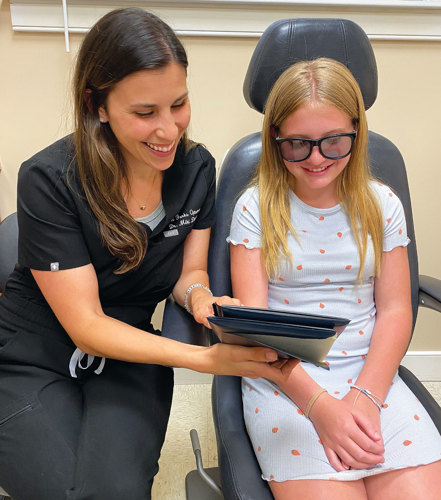
As eye and vision problems in infants can cause developmental delays, and one in four school-aged children has a vision disorder, according to the American Optometric Association, optometrists should be prepared to manage these young patients.
This two-part article discusses how to accomplish that, with part one providing advice on how to both prepare for and perform a pediatric eye exam. (See “The developing visual system,” below.)
The developing visual system
The visual system begins developing in utero in the earliest stages of pregnancy at about seven weeks gestation and continues to grow throughout the 40 weeks.
Upon birth, the eye itself is still not mature, with the eyeball being about 2/3rd of its full size, the muscles of the eye unmyelinated and uncoordinated, and an almost non-existent accommodative and binocular system.
With high-refractive error and steep corneas at birth, infants can only see about 20/400 or so.
As the child grows, the eye undergoes many changes during the process of emmetropization and requires proper input and stimulation to gather, process, and integrate visual input to develop normal binocular vision and eyesight.
How to prepare
Pediatric exam success is contingent on getting past the traditional adult exam. You’re dealing with a very different patient population here. Therefore, you want to be as flexible (literally and figuratively) as possible with these exams. My advice:
First, discuss with the parent or caregiver that the baby’s or child’s cooperation and attention will dictate the amount of data that you are able to glean, which may necessitate additional follow-up appointments for a full assessment.
Next you’ll want to begin the exam, prioritizing any concern the parent may have. For example, you may do binocular vision testing before visual acuity testing if the parent is there to get their child assessed for an eye turn.

You want to get out of the box of a traditional exam and garner as much information as possible, even if the order of the exam is not how you would typically perform it.
Additionally, get comfortable with the idea that you may have to perform retinoscopy or binocular indirect ophthalmoscopy while squatting on the floor or playing “peekaboo” with the child.
How to perform
Here are the specific action steps to follow:
• Acquire a case history. This is comprised of a detailed discussion of pregnancy, birth history, family history, and developmental milestones. Comprehending this early history is critical in understanding whether any interruptions to the child’s development occurred that affect their ocular health. For example, if a child was born prematurely, we know there are higher risks for refractive errors or strabismus.1
• Assess visual acuity through force preferential looking (FPL) techniques. When assessing acuity with FPL, ensure that the baby does not see you. The idea behind this technique is to understand that a baby will always look at “something” vs. “nothing.” If the gratings are blurred, the baby will look equal to the blank, gray plate or lose interest in the testing. Depending on the cards used, there will be a little peephole to look through, as you present the targets to the infant, or you can slyly peak over or between cards to watch the baby. Testing is done in a staircase method, with you observing whether the baby is looking/turning their head toward the appropriate target and you determining maximal VA. This can be done within three-to-five minutes clinically.2 To feel confident in this technique, before you begin, present the largest grating test plate or a toy to the baby, to understand their looking style (head turn, eye movement, gesture). This way, you’ll know what to look for.
As a child’s age and capabilities increase, the optometrist can gain more subjective information from them. I recommend considering single optotype picture matching for VAs for children who have not yet mastered their alphabet/numbers.
Additionally, I suggest performing both dry and wet retinoscopy to understand the patient’s visual system and refractive error.
• Evaluate the binocular system. This entails using cover testing or the Brückner/Hirschberg assessment, as well as nearpoint of convergence and stereopsis assessment, dry retinoscopy, and a full cycloplegic exam. I highly recommend getting a colorful, spinning or light-up toy to help keep the baby’s visual attention long enough to do these tests. For distance, most digital acuity charts play some sort of show or movie; worst-case scenario you can always have the parents use their phones to direct the baby’s visual attention in a certain gaze. As the baby gets older, they may try to grab the occluder; if that is the case, the OD can always use their hand to quickly perform a cover test.
• Conduct an ocular health assessment. For babies ages six months and older, 1% cyclopentolate is the preferred drop of choice to use for full cycloplegia to assess their refractive error and ocular health. This is due to cyclopentolate’s level of cycloplegic effect (more than tropicamide) and lower risks vs. atropine.3 For infants younger than six months of age, 0.2% to 0.5% of cyclopentolate is preferred.4,5
As administration of the cycloplegic agent may be difficult, I recommend spraying the lashes with the agent, as this works sufficiently.
Ocular health assessment can be done with either a direct ophthalmoscope or a binocular indirect ophthalmoscope and a 20 D lens. Peripheral views may be limited, but optic nerve head and macula observation are often facilitated with flash toys or making noises to have the infant look at you.
This initial exam serves as a baseline of the infant’s developing eye and to enable the observation of any large refractive errors, strabismus, or ocular health problems. Possible ocular health issues that may present include congenital cataracts, retinal issues or even pediatric glaucoma. If all findings are within normal, that child can be seen again between the ages of three to five and then annually thereafter.6 (See “InfantSee: an overview,” below.)
InfantSee: an overview
The American Optometric Association recommends that a child’s first eye exam is performed between six to 12 months of age through the organization’s InfantSEE program. This program provides a comprehensive eye exam at no cost to the patient, regardless of insurance status, with a certified provider. Visit infantsee.org.
More to come
In part two of this article, I discuss the OD’s role in treating the most prevalent pediatric conditions: refractive error, strabismus, amblyopia, cataract, retinoblastoma, retinopathy of prematurity, nystagmus, Stargardt disease, and color blindness. OM
References
1. Xie X, Wang Y, Zhao R, et al. Refractive status and optical components in premature infants with and without retinopathy of prematurity: A 4- to 5-year cohort study. Front Pediatr. 2022;:(10):922303. doi: 10.3389/fped.2022.922303.
2. McDonald MA, Dobson V, Sebris SL, Baitch L, Varner D, Teller DY. The acuity card procedure: a rapid test of infant acuity. Invest Ophthalmol Vis Sci. 1985;26(8):1158-62.
3. Major E, Dutson T, Moshirfar M. Cycloplegia in Children: An Optometrist’s Perspective. Clin Optom (Auckl). 2020; (12): 129–133. doi: 10.2147/OPTO.S217645.
4. American Optometric Association: Guideline Brief 2017: Evidence-Based Clinical Practice Guideline: Comprehensive Pediatric Eye and Vision Examination. Accessed August 11, 2024. bit.ly/AOAPediatricEyeandVisionExam
5. Wickim SM, Amos JF. Chapter 21: Cycloplegic refraction. In Bartlett JD, Jaanus SD, eds. Clinical Ocular Pharmacology, 5th edition. St. Louis: Butterworth-Heinemann; 2008; 343-48.
6. AOA. Infant See: About Us. Accessed July 3, 2024. InfantSEE. https://www.infantsee.org/about-us?sso=y.


