This is the first in a series focusing on the diagnosis and treatment of geographic atrophy.
Geographic atrophy (GA) is a form of advanced dry age-related macular degeneration (AMD). As retinal cells die, irreversible damage occurs, leading to vision loss that cannot be restored. Early detection and monitoring of GA progression are crucial for optometrists (ODs) to proactively address impending threats to a patient’s vision.
It is important to note that visual acuity is not always well- correlated with GA progression. Therefore, ODs must rely on dilated fundus exams and multimodal imaging to identify and monitor GA lesions, which grow over time and lead to significant visual impairment.
Lesion Characteristics and Detection
GA lesions present as well-demarcated areas of retinal atrophy. These lesions typically appear as a pale, depigmented region in the macula and can vary in size and shape. The borders are distinct, often surrounded by a rim of hyperpigmentation or atrophic changes.
Imaging techniques such as fundus autofluorescence (FAF) and optical coherence tomography (OCT) are valuable for visualizing and assessing GA lesions. FAF imaging is particularly useful for detecting the early stages of GA, as it highlights areas of decreased autofluorescence corresponding to the atrophic lesions. OCT allows for a more detailed view of the retinal layers and helps to assess the extent of tissue loss and damage in the retina. These images provide critical insight into disease progression and aid in identifying risk factors for faster progression.
Four Key Indicators of Faster GA Progression
While the progression of GA varies among patients, certain characteristics can suggest a higher risk of faster disease advancement:
- Multifocal lesions: The presence of multiple GA lesions in one or both eyes is a significant risk factor for faster progression (see Figure 1).
- Extrafoveal lesions: Lesions located outside the fovea, but still within the macula, are more concerning than those confined to the central fovea (see Figure 2).
- Bilateral lesions: The presence of GA in both eyes significantly raises the likelihood of faster progression (see Figure 3).
- Hyperfluorescence on imaging: Particularly on FAF imaging, this can indicate areas of active degeneration (see Figure 4).
Geographic atrophy is a progressive, irreversible condition that requires timely detection and intervention. Optometrists have the unique opportunity to detect GA early through careful examination and imaging and identify risk factors for faster progression. Because of this, ODs are the cornerstone of early detection that can help their patients optimize the opportunity to slow progression by referring for treatment.
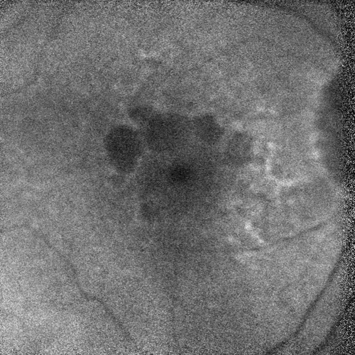
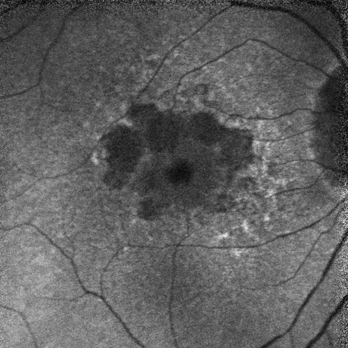
Figures 1A and 1B. Multifocal lesions often indicate a more aggressive form of the disease.
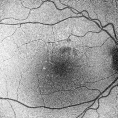
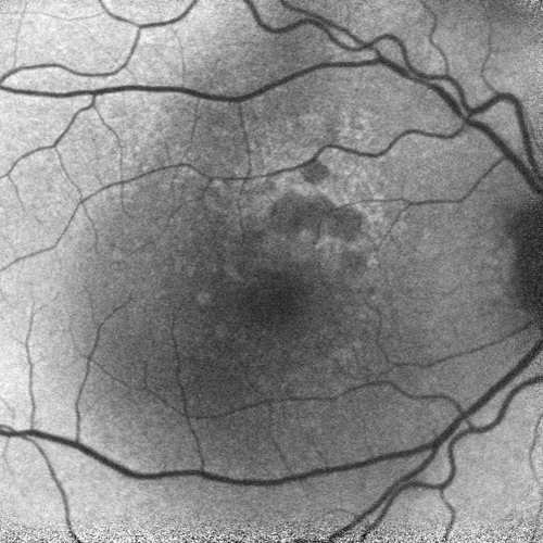
Figures 2A and 2B. Extrafoveal lesions often grow faster than foveal lesions.
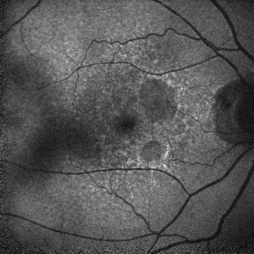
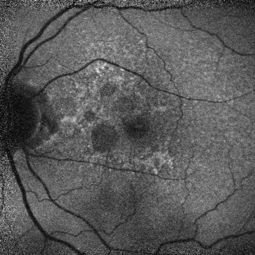
Figures 3A and 3B. 5/3/16
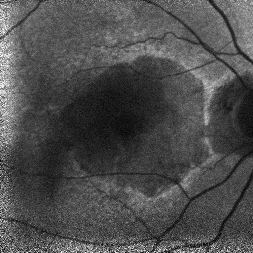
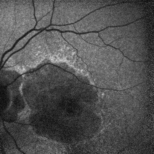
Figures 3C and 3D. 2/25/21
Figures 3A, 3B, 3C, and 3D. Bilateral GA progression often reflects the systemic nature of AMD.
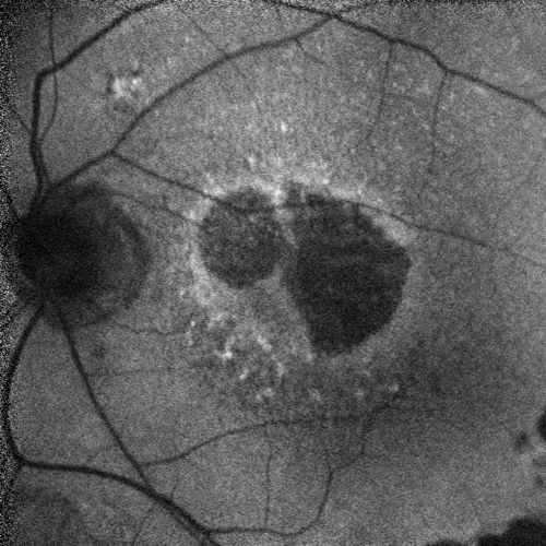
Figure 4. Hyperfluorescence indicates excess lipofuscin in the retina. The banded pattern suggests that there are damaged retinal cells.
This editorially independent content is sponsored by 


