


While working in a multi-specialty clinic, there are plenty of opportunities to experience new technology in imaging the cornea and the anterior segment. However, a common theme with new technology is that as the device becomes more specialized, so does the patient population. This can create a barrier to return on investment for interested providers—will there be enough reasons to use this device in my practice?
One device that has passed this test in our practice is our Anterion (Heidelberg Engineering), an anterior segment swept source OCT. While it was originally selected for mapping the epithelium of the cornea, it has turned into a workhorse instrument when evaluating patients with keratoplasties, scleral lenses, before and after refractive surgery, as well as many other conditions.
Corneal Transplant Management
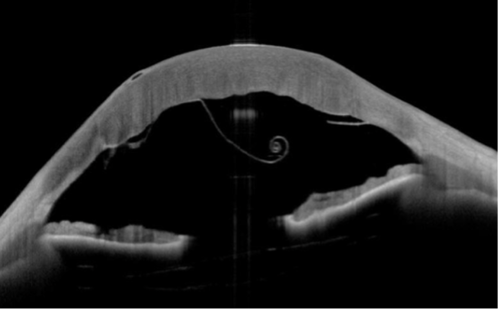
The Anterion is a great tool to evaluate and educate our patients who have undergone endothelial keratoplasties. The imaging module displays the position of the transplanted endothelial layer to the host cornea and easily images through corneal haze in a case of moderate corneal swelling. These detailed images assist in the decision to rebubble or reposition graft tissue. For complex corneal imaging, there are straight-forward cues to guide the provider or technician, ensuring an efficient patient experience and high-quality image.
Precision in Scleral Lens Prescribing
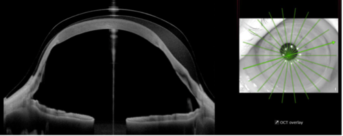
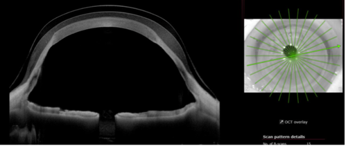
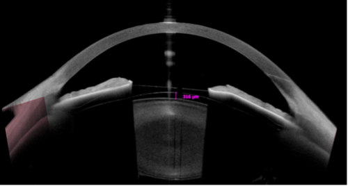
AS-OCT has been utilized in scleral lens prescribing for a long time; however, without being able to image the entire lens in one scan, it is difficult to build an accurate assessment of the cornea-lens relationship, and images can be misleading. On this AS-OCT, a customized scleral lens imaging setting can scan about 10 images at a 16 mm chord across the lens in about 5 seconds, allowing for accurate assessment of lens position, central vault, and limbal clearance. These scans are particularly helpful when evaluating corneas with complex shapes, such as a tilted graft, as shown in the images below. A 360-degree scan over the limbus allows for precise limbal clearance and mid-peripheral curvature prescribing to create an intricate scleral lens chamber for this asymmetric cornea shape.
Refractive Surgery Management
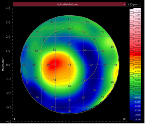
When screening patients for their candidacy for LASIK, epithelial mapping can assist in identifying early corneal ectasias. The epithelial layer can mask the presence of mild keratoconus, as the tissue thickens at the base of the cone and thins at the apex. This effectively minimizes the true shape of the corneal elevation, and the diagnosis can be missed when imaged with corneal topography.
The Anterion is also very effective when planning ICL surgery for high myopes. The metrics module can image the anterior chamber depth, corneal white-to-white, keratometry, pupil size, corneal pachymetry, and other anterior segment measurements. Post-operatively, the anterior segment can be imaged to assess for change over time, including findings like the ICL vault, iris shape, and anterior chamber angle changes.
As the Anterion gets more utilization in our practice, our technician team has learned a significant amount about anterior segment anatomy and post-surgery findings. This has elevated the provider efficiency and patient care and satisfaction.


This editorial content was supported via unrestricted sponsorship