


“Form follows function” may be a workable principle when employed by a design architect, but an optometrist must often rehabilitate function when form has suffered a change or loss. When it comes to corneal architecture, visual function is at the complete mercy of the structural form. As the stroma warps, the optics rebel. When the epithelium compensates, it does so imperfectly. And when herpes simplex virus leaves its indelible fingerprint, the corneal form can transform from the eye’s perfect window into something closer to frosted glass.
In a busy anterior segment practice, cases like this appear with uncanny regularity: complex, scarred, asymmetric corneas that make us appreciate—sometimes grudgingly—the sheer importance of detailed corneal mapping. Without it, we are flying blind. With it, we have the chance to turn hopeless cases into triumphs.
Case Presentation: A Scarred and Stubborn Cornea
A 27-year-old female patient with a history of recurrent herpes simplex keratitis OS was referred after her vision plateaued at 20/100 despite multiple attempts at contact lens rehabilitation. The referring ophthalmologist, facing therapeutic exhaustion, warned that if a lens could not provide functional vision, a corneal transplant would be the next step—though he admitted reluctance given her youth.
On exam:
- Anterior segment: diffuse, irregular stromal scarring with non-active stromal vessels; intact epithelium.
- Other findings: unremarkable.
The clinical dilemma was clear. The scarring had profoundly altered corneal architecture. The failed lens fittings made sense—but why? And could technology illuminate a path to rehabilitation short of transplantation?
Anterion at the Crossroads of Form and Function
Enter the Anterion (Heidelberg Engineering), armed with its Cornea and Imaging Apps. These tools are, in many ways, the optometrist’s equivalent of MRI in neurology: fast, high-resolution, and brutally honest about structural detail.
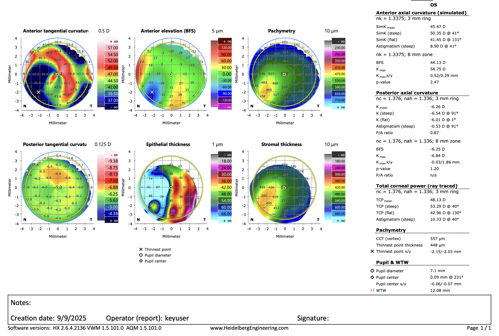
- Anterior tangential curvature map: revealed a corneal landscape resembling an irregular mountain range more than an optical surface. Distortion was global, explaining the contact lens failures.
- Anterior elevation map: confirmed chaotic height variations—a cartographer’s nightmare.
- Pachymetry map & stromal thickness map: showed severe irregularity, with stromal fibrosis driving local stromal thinning and epithelial thickening.
- Epithelial thickness map: revealed compensatory thickening over areas of stromal loss, a well-documented adaptive response but one that further compromises smooth optics.
The verdict was clear: this cornea was not only scarred, it was also architecturally untrustworthy. A conventional lens would be doomed to fail. The form dictated a custom scleral lens.
Treatment Outcome: Vision Restored
A custom scleral lens was designed using the Anterion data. On fitting, the patient achieved 20/20-2 best-corrected visual acuity. The transplant was averted. In the end, advanced imaging transformed a “last-ditch” referral into a story of successful rehabilitation.
The Imaging App: A Second Act
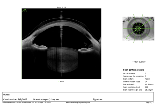
During fitting, the Imaging App proved equally invaluable:
- Simultaneous multi-meridian scans (5 meridians at once): allowed comprehensive evaluation of lens–cornea–sclera interaction.
- Extended 16.5-mm scan diameter: offered true limbus-to-sclera coverage, critical in scleral lens cases.
- Swept-source OCT with superior depth and resolution: cut cleanly through opaque scar tissue, visualizing both density variations in fibrosis and the associated epithelial hypertrophy.
This meant that lens clearance, scleral landing, and corneal–epithelial responses could all be monitored in exquisite detail, minimizing trial-and-error and expediting the path to success.
Lessons From the Case
- Corneal form dictates visual function. Distorted structure = distorted optics. Every map confirms this.
- Epithelial compensation is clever but imperfect. Thickness changes may stabilize shape, but they create new irregularities.
- Technology matters. Instruments like the Anterion allow us to cut through the clinical fog—literally through scar tissue—and see what’s happening beneath.
- Transplants can sometimes wait. With the right diagnostic data, even eyes that look destined for keratoplasty can achieve remarkable vision with specialty contact lenses.
Conclusion
The cornea is a structure that writes its own destiny: When its form changes, so too does vision. Modern anterior segment imaging doesn’t just show us the damage—it shows us the roadmap back to function.
In this case, the Anterion helped reveal the “why” behind repeated lens failures, guided a successful scleral lens fit, and spared a young woman a transplant. The message for the cornea specialist is clear: If form predicts function, then understanding form with precision is the surest way to restore function.
Sometimes, the best scalpel is a map.

This editorial content was supported via unrestricted sponsorship