


A cross-sectional analysis of more than 45,000 eyes from the UK Biobank and an independent cohort of young adults in Scotland has introduced a novel biomarker—fundus refraction offset (FRO)—to quantify individualized anatomic variations in myopia.
Published in JAMA Ophthalmology, the study proposes FRO as a deep learning (DL)–derived metric that captures discrepancies between fundus appearance and spherical equivalent refraction (SER), offering a refined approach for assessing posterior segment severity and potential myopic complications “even among individuals with similar SER or axial length (AL) and demographic characteristics,” wrote the authors, led by Fabian Yii, BSc, of The University of Edinburgh.
Current clinical assessments of myopia primarily rely on SER and AL, which provide one-dimensional, on-axis measurements of the eye. These metrics often fail to capture subtle, off-axis anatomic differences. For example, 2 patients with identical SER may show markedly different retinal morphology and risk profiles for myopic degeneration. FRO can address this gap, the researchers explained.
FRO represents the deviation between actual SER and fundus-predicted SER—termed fundus equivalent refraction (FER). A negative FRO indicates a relatively more myopic-appearing fundus than expected for a given SER, whereas a positive FRO suggests a more hyperopic appearance.
In this study, a ResNet-18 DL model was trained on 31,670 images from healthy eyes in the UK Biobank to predict SER. The model was applied to a 30% holdout subset from the UK Biobank (internal unseen set) and a Caledonian student cohort (external data set). The model achieved strong correlations between FER and true SER across the training (r=0.97), internal (r=0.94), and external (r=0.78) data sets.
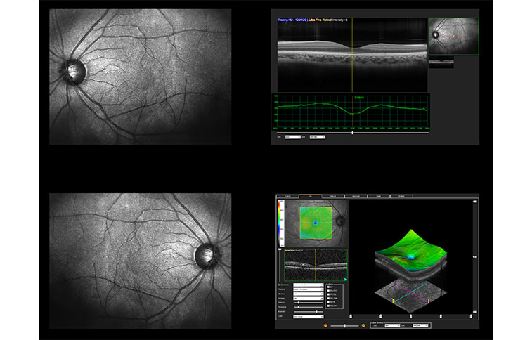
The researchers found that FRO was significantly associated with anatomic differences that were observable on OCT imaging, independent of SER or AL:
- Macular Thickness (MT):
- UK Biobank: For each −1.00 D increase in FRO, MT decreased by 0.64 μm (P<.001).
- Caledonian cohort: MT reduction of 2.09 to 2.45 μm (P=.02–.008) depending on model adjustments.
- Choroidal Vascularity Index (CVI):
- Higher CVI was associated with more positive FRO (P<.001), indicating better choroidal perfusion.
- No association was found between FRO and choroidal area (P>.30 in all models).
These findings held true even after adjusting for age, sex, race, and on-axis measurements (SER or AL).
FRO may aid in identifying eyes that are at higher risk of myopia-related complications despite having similar SER or AL values. Because it leverages fundus image data which are routinely acquired in practice, it could be integrated into screening tools or longitudinal risk assessments.
The authors compared FRO to "retinal age gap"—a DL-derived metric that predicts biologic aging of the retina and correlates with systemic disease risk. FRO might also predict future axial elongation or myopia onset and help to target preventive interventions in susceptible patients.
The researchers acknowledged limitations, including varied performance among imaging systems and a homogenous cohort of patients that skewed heavily White and middle-aged, which may limit generalizability. They recommended longitudinal studies to assess whether FRO can independently predict myopic progression or complications.
“A promising future direction,” the authors concluded, “is to explore whether baseline FRO is independently associated with the risks of future myopic complications, beyond the influence of baseline myopia severity and other potential covariates. Similarly, investigating whether a relatively more myopic-looking (or less hyperopic-looking) fundus—reflected by a more negative (or positive) FRO—signifies that the eye concerned is already on a trajectory toward rapid myopia progression (or myopia onset) is another promising direction for future research.”