


Researchers affiliated with the National Institutes of Health have developed a three-dimensional “digital twin” of retinal pigment epithelium (RPE) cells, creating a platform to study how cellular organization changes in age-related macular degeneration (AMD).
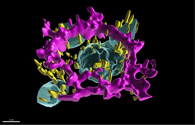
The model is based on induced pluripotent stem cell–derived RPE generated at the National Eye Institute (NEI), using 3D imaging data from approximately 1.3 million cells collected across nearly 4,000 fields of view. Investigators trained an artificial intelligence algorithm, called POLARIS (Polarity Organization with Learning-based Analysis for RPE Image Segmentation), to identify nuclei, mitochondria, cytoskeletal structures, and overall cell morphology at subcellular resolution.
RPE cells require apical-basal polarity to recycle photoreceptor outer segments and manage nutrient and waste exchange. The team quantified changes in cell shape, organelle distribution, and spatial organization across developmental stages, finding that healthy cells follow a predictable trajectory toward polarization.
The resulting atlas distinguishes polarized and nonpolarized states, providing a reference for examining how polarity is disrupted in AMD. According to NEI Scientific Director Kapil Bharti, PhD, the approach may support therapeutic discovery and could be adapted to other diseases involving loss of cellular organization.
"By combining AI with mathematical modeling, we've created a window into cellular processes that were previously hidden from view," said NEI research fellow Davide Ortolan, PhD, the senior leader on this program. "This technology doesn't just help us understand what's happening in AMD, it gives us a platform to discover how to fix it."
The study was published February 6 in Nature Partner Journal-AI.