Access, ability, and association aid O.D.s
OCT-angiography (OCT-A) provides complimentary and supportive information when it comes to our glaucoma patients. These benefits of OCT-A may be summarized by the following additional A’s.
1 ACCESS TO DEEPER LAYERS…
By way of analogy, a tree is subject to external pressures — such as wind or heat — which, if extreme, could cause damage to the tree, commonly starting with the branches. There are other factors — such as poor water and nutrient supply — which, if extreme, could also cause damage to the tree, commonly starting with the roots. Although the damage to the branches may be more readily noticeable, damage to the nourishing root supply is not as noticeable and may precede visible damage by several years.
Similar to a tree, the optic nerve is subject to IOP forces which, if significant (and for a prolonged period), could cause irreversible glaucomatous optic neuropathy. Additionally, there are vascular dysregulation factors (particularly within the microcirculation of the posterior ciliary artery), which, if extreme, could impair the otherwise nourishing vascular optic nerve supply.1
Therefore, to arrive at a complete picture of optic nerve health, and similar to a tree, we need to go deeper… We need a sort of noninvasive vascular root system analysis. OCT-A provides us with access to deeper layers into the optic nerve and choroidal vasculature. Because of this deeper access with OCT-A, studies show that these abnormal autoregulation responses seem to occur in all stages of glaucoma (from early to late progression) and that these functional and morphological changes may occur independently of the IOP levels.2
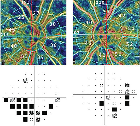
OD vs. OS

2 ABILITY TO DIAGNOSE EARLIER AND DETECT PROGRESSION LATER…
As abnormal autoregulation has been detected in all stages of glaucoma, we should not be surprised by studies dating back nearly a decade that report decreased optic nerve perfusion occurring even in pre-perimetric glaucomatous eyes when compared to normal eyes.3 At the other end of the glaucoma disease spectrum, OCT-A provides us with the ability to track glaucomatous progression later, even in advanced glaucoma due to lower measurement floors.4 Daneshvar et al. conclude from their review that “almost all published articles demonstrate a significant reduction of blood flow, capillary diameter and vascular density in glaucomatous eyes…[and that]…these differences were detectable even in glaucoma suspects and eyes that had pre-perimetric glaucoma and increased proportional to the severity of glaucoma damage.”5
3 ASSOCIATION WITH STRUCTURAL AND FUNCTIONAL DAMAGE…
Throughout the spectrum of glaucoma disease severity, capillary dropout is strongly and spatially associated with RNFL defects as well as correlating VF defects.5,6 In short, such growing clinical correlation adds increased certainty to our diagnosis as well as added guidance to our future testing and treatment options.
4 A FINAL THOUGHT…
Diagnosing glaucoma and monitoring for its progression can sometimes seem like an ever-changing puzzle, but OCT-A testing may provide the additional pieces to complete the clinical picture. As such, and even if OCT-A testing may not be readily available in all our clinics, we should consider reaching out to other colleagues within our area with whom we can, perhaps, partner and share in this technology on behalf of our patients. OM
REFERENCES
- Jia Y, Tan O, Tokayer J, Potsaid B, et al. Split-spectrum amplitude-decorrelation angiography with optical coherence tomography. Opt Express. 2012;20:4710–4725. doi: 10.1364/OE.20.004710
- Wareham LK, Calkins DJ. The Neurovascular Unit in Glaucomatous Neurodegeneration. Front Cell Dev Biol. 2020;8:452. doi: 10.3389/fcell.2020.00452. eCollection 2020.
- Jia Y, Tan O, Tokayer J, Potsaid B, Wang Y, Liu JJ, et al. Split-spectrum amplitude-decorrelation angiography with optical coherence tomography. Opt Express. 2012;20:4710–4725. doi: 10.1364/OE.20.004710.
- Van Melkebeke L, Barbosa-Breda J, Huygens M, Stalmans I. Optical Coherence Tomography Angiography in Glaucoma: A Review. Ophthalmic Res. 2018;60(3):139-151. doi: 10.1159/000488495.
- Daneshvar R, Nouri-Mahdavi K. Optical Coherence Tomography Angiography: A New Tool in Glaucoma Diagnostics and Research. J Ophthalmic Vis Res. 2017;12(3):325-332. doi: 10.4103/jovr.jovr_36_17.
- Holló G. Optical Coherence Tomography Angiography in Glaucoma. Turk J Ophthalmol. 2018;48(4):196-201. doi: 10.4274/tjo.53179.


