The incidence of AMD is dramatically increasing. It is predicted to affect up to 288 million people worldwide by 2040.1 The non-neovascular or “dry” form is classically distinguished from the neovascular or “wet” type based upon pathology, time course and severity.1,2 We know that dry AMD may eventually progress or “convert” to the wet form, but we also have patients who appear to be already “wet at the onset.” In these cases, neovascular AMD (nAMD) emerges acutely and rapidly progresses to severe central vision loss.3 In many cases, the dry form stays dry, but progresses to its end stage of geographic atrophy (GA), eventually impairing visual function.4
We also know that oxidative stress, complement dysregulation and hypoxia all contribute to AMD’s pathogenesis, but the specifics of how each one contributes remain elusive.5 Are wet and dry AMD two different diseases, or do they truly share common pathogenic mechanisms? If so, which mechanisms underlie the two forms? Are they different for the two variants, or do they stem from common metabolic alterations?
The pathology of AMD features the formation of drusen, subretinal drusenoid deposits or both, along with disturbances of the retinal pigment epithelium (RPE) and Bruch’s membrane.3 Depending on the phenotypic variant, the proliferation of new blood vessels may ensue. This emergence of macular neovascularization (MNV) represents the key distinguishing point between dry and wet AMD.3,4
Despite this distinction, an overlap exists both in the phenotypes and the biochemical mechanisms underlying these seemingly disparate clinical conditions.5 In fact, wet AMD often occurs on a background of dry AMD, and both forms may occur in the same eye at different stages of the disease. This is consistent with observations suggesting that GA and MNV are different though interconnected manifestations of the same disease.5
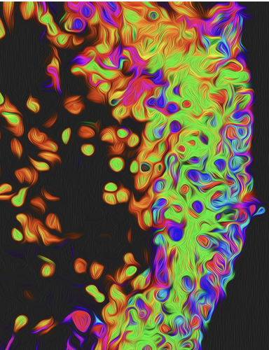
Researchers from the Wilmer Eye Institute sought to identify key molecular events that contribute to the development of both MNV and GA. They used a combination of cell-based models, human-induced pluripotent stem cell–derived RPE and retinal organoids, as well as animal models for ocular oxidative stress (Figure 1).6 The team demonstrated that oxidative stress and hypoxia synergistically promote the accumulation of hypoxia-inducible factor (HIF-1α) in RPE and photoreceptors. They found that this factor contributed to the development of MNV.6
The Wilmer team also found that increased HIF-1α in photoreceptors exposed to oxidative stress reduced cell death, an early event in the development of GA.6 These results suggest that in patients with AMD, increased expression of HIF-1α in RPE exposed to oxidative stress promotes the development of MNV, but inadequate HIF-1α expression in photoreceptors contributes to the development of GA.6
These observations provide a molecular explanation for how oxidative stress can simultaneously influence whether a patient with AMD develops MNV or GA. Therefore, modulation of HIF-1α may be an effective therapeutic approach to prevent progression to advanced AMD (defined as either GA or MNV).
Key Clinical Takeaways
- AMD is classically divided into the dry and wet forms.
- The development of macular neovascularization (MNV) represents the key distinguishing point between dry and wet AMD.
- The level of hypoxia-inducible factor (HIF-1α) accumulation may play a central role in determining whether patients with AMD remain stable, progress to MNV or develop GA.
- A careful balance of HIF-1α levels must be maintained to prevent vision loss in the eyes of patients with AMD.
Reference(s):
- Wong WL, Su X, Li X, Cheung CMG, Klein R, Cheng C-Y, Wong TY. Global prevalence of age-related macular degeneration and disease burden projection for 2020 and 2040: a systematic review and meta-analysis. Lancet Glob Health. 2014 Feb;2(2):e106–e116. doi:10.1016/S2214-109X(13)70145-1
- Jager RD, Mieler WF, Miller JW. Age-related macular degeneration. N Engl J Med. 2008 Jun;358(24):2606–2617. doi:10.1056/NEJMra0801537
- Gehrs KM, Anderson DH, Johnson LV Hageman GS. Age-related macular degeneration--emerging pathogenetic and therapeutic concepts. Ann Med. 2006;38(7):450–471. doi:10.1080/07853890600946724
- Girmens J-F, Sahel J-A, Marazova K. Dry age-related macular degeneration: a currently unmet clinical need. Intractable Rare Dis Res. 2012 Aug;1(3):103–114. doi:10.5582/irdr.2012.v1.3.103
- Pinelli R, Biagioni F, Limanaqi F, Bertelli M, Scaffidi E, Polzella M, Busceti CL, Fornai F. A re-appraisal of pathogenic mechanisms bridging wet and dry age-related macular degeneration leads to reconsider a role for phytochemicals. Int J Mol Sci. 2020 Aug;21(15):5563. doi:10.3390/ijms21155563
- Babapoor-Farookhran S, Flores-Bellver M, Niu Y, et al. Pathologic vs. protective roles of hypoxia-inducible factor 1 in RPE and photoreceptors in wet vs. dry age-related macular degeneration. PNAS. 2023 Dec;120(50): e2302845120. https://www.pnas.org/doi/10.1073/pnas.2302845120


