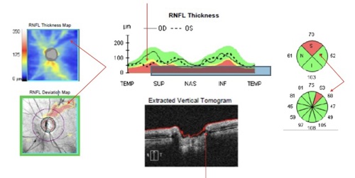
At the lecture “OCT Interpretation of Optic Nerve Head and Retinal Nerve Fiber Layer Scans,” which occurred yesterday at 9 a.m., Lee Vien, OD, FAAO, and David Yang, OD, FAAO, discussed the crucial role of optical coherence tomography (OCT) in diagnosing and managing glaucoma.
“OCT is a powerful diagnostic tool in ocular disease management and clinicians should maximize the utility of the instrument to fully understand the information presented,” said Dr. Vien of the lecture.
Filling in gaps
The lecture addressed existing knowledge gaps in how to best utilize and interpret OCT results, with a particular focus on the optic nerve head and retinal nerve fiber layer (RNFL). For example, recognizing common OCT artifacts, such segmentation errors, were discussed.
Tackling technologies
Additionally, the lecture covered various OCT technologies, including time-domain, spectral-domain, and swept-source OCT, with explanations on how to use enhanced-depth imaging.
Further, attendees were guided through key anatomical features visible on OCT, such as optic disc margins, the neuroretinal rim, and RNFL bundles, and instructed on how to interpret OCT printouts, including thickness and deviation maps, tomograms, and optic nerve parameters.
Clinical situations
Case studies during the lecture showcased the use of OCT in various clinical situations, including preperimetric glaucoma, normal-tension glaucoma, and in glaucoma suspects with unique anatomical variations, such as situs inversus or high myopia. What’s more, each case emphasized how OCT findings, such as RNFL distribution and neuroretinal rim thickness, could guide diagnostic decisions and glaucoma management. OM
Advice for Academy attendees
"Take advantage of all that AAO has to offer," recommended Dr. Vien. "Although the annual AAO meeting is known for all CE all the time, there are numerous other activities, including school receptions, multiple networking events, and a large exhibit hall."


