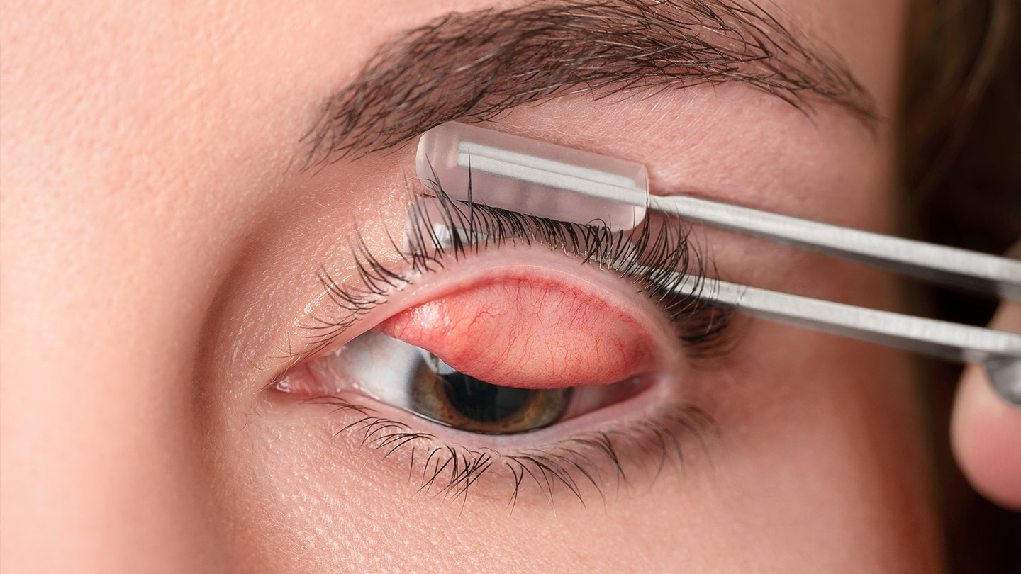
A 2025 review demonstrates that upper lid eversion should be considered an essential component of a comprehensive ocular examination.1 The reason: Failure to examine the superior palpebral conjunctiva may lead to overlooked diagnoses of ocular and systemic conditions, resulting in delayed intervention and less favorable patient outcomes. These conditions:
Meibomian Gland Dysfunction
Several studies underscore the clinical importance of assessing the upper lids to complete a comprehensive evaluation of the meibomian glands.1,2-8 Optometrists should consider evaluating the lids of contact lens wearers for meibomian gland dysfunction (MGD) because of its association with dissatisfied contact lens wearers.5,9 Additionally, contact lens dropout increases in patients who have worsening grading of upper lid meibomian gland tortuosity and plugging and meibum quality.5,9
Further, evaluating the upper lid in both contact and non-contact lens wearers can aid ODs in identifying patients who have MGD prior to and after cataract surgery, and secondary to systemic issues, such as Sjögren syndrome, systemic lupus erythematous, or rheumatoid arthritis.1
Lid Wiper Epitheliopathy
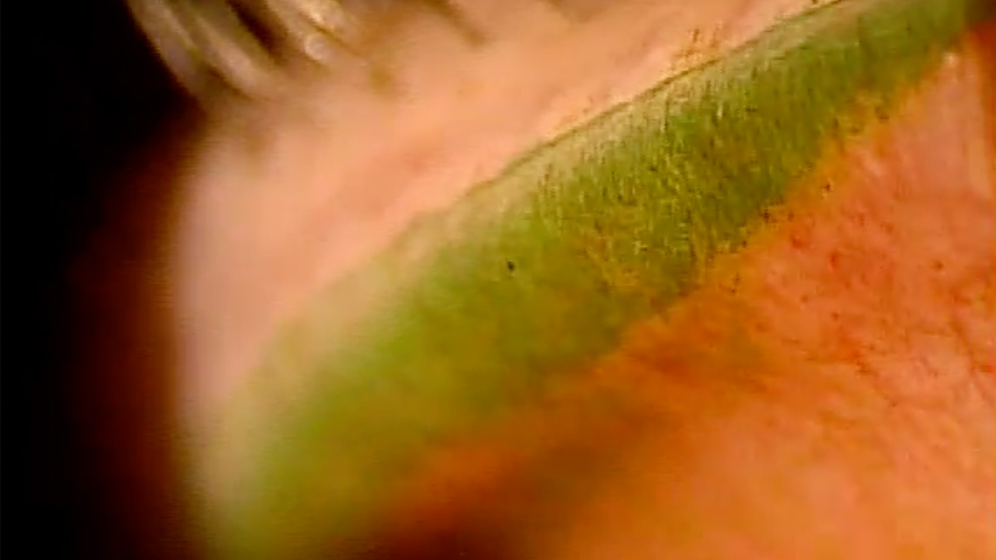
Upper lid eversion can also uncover lid wiper epitheliopathy (LWE), which appears as vital staining of the lid margin, believed to occur from increased friction among the palpebral lid and the opposing ocular surface structures.10 The condition is commonly associated with dry eye disease and ocular dryness symptoms in both contact and non-contact lens wearers.11,12 Underutilization of upper lid eversion can lead to missed diagnoses of LWE.13
Conjunctival Issues
Inflammation, foreign bodies, and concretionscan be identified via upper lid eversion. For example, ODs can easily observe subtle cases of giant papillary conjunctivitis by everting the upper lid and examining the palpebral conjunctiva in patients who present complaining of discomfort with contact lens wear.
Upper Lid Eversion in Clinical Evaluation
Assessment of the superior palpebral conjunctiva is an important component of a thorough ocular surface examination. Upper lid eversion allows clinicians to visualize structures that may not be apparent during standard anterior segment evaluation. As demonstrated above, routine examination of this area can aid in identifying ocular surface findings relevant to diagnosis and management.¹ OM
References
1. Gupta PK, Karpecki P. Comprehensive assessment of the meibomian glands by meibography: Why the upper eyelids matter. Cornea. 2025;44(1):128-135. doi:10.1097/ICO.0000000000003729
2. McCann LC, Tomlinson A, Pearce EI, Diaper C. Tear and meibomian gland function in blepharitis and normals. Eye Contact Lens. 2009;35(4):203-208. doi:10.1097/ICL.0b013e3181a9d79d
3. Eom Y, Choi KE, Kang SY, Lee HK, Kim HM, Song JS. Comparison of meibomian gland loss and expressed meibum grade between the upper and lower eyelids in patients with obstructive meibomian gland dysfunction. Cornea. 2014;33(5):448-452. doi:10.1097/ICO.0000000000000092
4. Pucker AD, Jones-Jordan LA, Marx S, et al. Clinical factors associated with contact lens dropout. Cont Lens Anterior Eye. 2019;42(3):318-324. doi:10.1016/j.clae.2018.12.002
5. Dogan AS, Kosker M, Arslan N, Gurdal C. Interexaminer reliability of meibography: upper or lower eyelid? Eye Contact Lens. 2018;44(2):113-117. doi:10.1097/ICL.0000000000000307
6. Maskin SL, Testa WR. Growth of meibomian gland tissue after intraductal meibomian gland probing in patients with obstructive meibomian gland dysfunction. Br J Ophthalmol. 2018;102(1):59-68. doi:10.1136/bjophthalmol-2016-310097
7. Maskin SL, Testa WR. Infrared video meibography of lower lid meibomian glands shows easily distorted glands: implications for longitudinal assessment of atrophy or growth using lower lid meibography. Cornea. 2018;37(10):1279-1286. doi:10.1097/ICO.0000000000001710
8. Pucker AD, Jones-Jordan LA, Kunnen CME, et al. Impact of meibomian gland width on successful contact lens use. Cont Lens Anterior Eye. 2019;42(6):646-651. doi:10.1016/j.clae.2019.06.004
9. Gao Y, Huang M, Song W, Li Y, Yan X. Lid wiper epitheliopathy: an early sign of dry eye diagnosis. Front Med (Lausanne). 2025;12:1593430. Published 2025 Jun 5. doi:10.3389/fmed.2025.1593430
10. Daniel E, Pistilli M, Ying GS, et al. Association of meibomian gland morphology with symptoms and signs of dry eye disease in the Dry Eye Assessment and Management (DREAM) study. Ocul Surf. 2020;18(4):761-769. doi:10.1016/j.jtos.2020.07.014
11. Yeniad B, Beginoglu M, Bilgin LK. Lid-wiper epitheliopathy in contact lens users and patients with dry eye. Eye Contact Lens. 2010;36(3):140-143. doi:10.1097/ICL.0b013e3181d94e82
12. Allen RC. Eversion of the upper eyelid. Webeye.ophth.uiowa.edu. January 12, 2017. Accessed September 18, 2025. https://webeye.ophth.uiowa.edu/eyeforum/atlas-video/eversion-of-upper-eyelid.htm#gsc.tab=0
13. Korb DR, Greiner JV, Herman JP, et al. Lid wiper epitheliopathy and dry eye symptoms. Eye Contact Lens. 2005;31(5):235‑239. doi:10.1097/01.ICL.0000186137.93693.4f