


A 56-year-old Haitian female patient presented with complaints of bilateral blurred vision. Her medical history was significant for sickle cell trait, hypertension, hypercholesterolemia, and diabetes mellitus of unknown duration. Her most recent blood glucose level was 180 mg/dL, with a hemoglobin A1c of 11%. Entrance testing was unremarkable. Best-corrected visual acuity measured 20/20 in the right eye (OD) and 20/400 in the left eye (OS). Slit-lamp biomicroscopy revealed mild cortical lens changes bilaterally. There was no evidence of iris neovascularization, and gonioscopy was negative for neovascularization of the angle. Intraocular pressure measured 20 mmHg OD and 22 mmHg OS by Goldmann applanation tonometry.
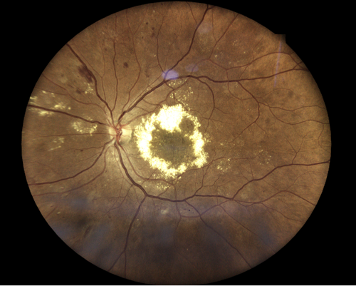
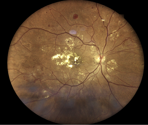
Fundus examination revealed pink, well-defined optic nerves bilaterally without evidence of neovascularization of the disc. There was extensive lipid exudation with associated macular edema in both eyes, along with numerous dot-and-blot and flame-shaped hemorrhages involving the posterior pole and extending into the equator bilaterally. The retinal arterioles appeared attenuated with arteriovenous crossing changes and an increased arteriolar light reflex, and findings were consistent with hypertensive and arteriosclerotic changes.
Additionally, diffuse exudation was noted throughout the equator, with a coinciding orangish-pink retinal hemorrhage observed superiorly in the right eye. Areas of anomalous retinal vasculature were identified in the inferotemporal and superonasal quadrants of the right eye. Overall, the clinical presentation was suggestive of a multifactorial retinopathy, most consistent with diabetic retinopathy with contributory sickle cell retinopathy.
Optical coherence tomography (OCT) and OCT angiography (OCT-A) were performed using the Optovue Solix Spectral Domain OCT system (Visionix) to assess for the presence of neovascularization and macular edema. Due to the equatorial location of the anomalous vasculature, 9x9 mm and 12x12 mm OCT-A were selected.
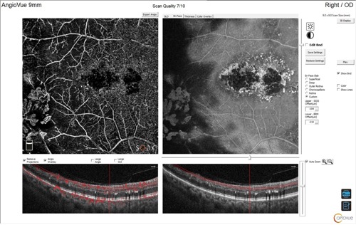
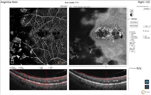
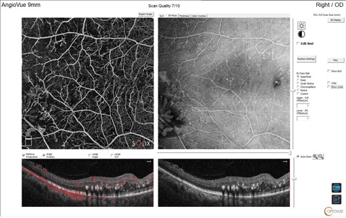
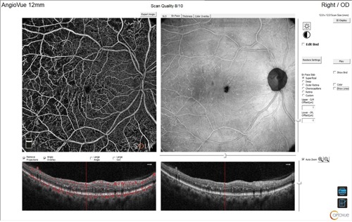
The utilization of OCT-A was instrumental in confirming the diagnosis of proliferative diabetic and sickle cell retinopathy and identifying associated macular edema. Employing advanced imaging technologies enhances diagnostic accuracy and supports improved clinical decision-making.

This content is sponsored by Visionix