


“Since using the Biovance 3L Ocular, I have found that the comfort is really good for patients,” he says. “And because it is decellularized, it takes out the donor DNA and any potential inflammatory cells—reducing the risk of having any autoimmune or inflammatory reaction.”
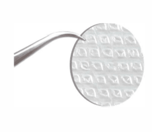
Biovance 3L Ocular is a trilayer decellularized basement membrane (DBM) used in ocular surgery and in OSD applications that include corneal and conjunctival-related injuries or defects such as persistent epithelial defects, corneal epithelial defect, pterygium repair, fornix reconstruction, and other procedures.
Like Dr. Gerson, Natalie Noble, OD, of Noble Vision Center in Greensburg, Pennsylvania, and Thomas M. Chester, OD, FAAO, of Cleveland Eye Clinic in Ohio, agree that the Biovance 3L Ocular offers patient and practice value.
Patient Value
What Dr. Noble likes the most about this membrane is “the improvement in corneal integrity to improve signaling so that all of the tear layers can improve in function.” She also likes the benefit of a DBM. “Not only is it giving the cornea all of this great cellular matrix and epithelial healing, but it also actually prevents some of the inflammatory response.”
For Dr. Chester, comfort for patients is also essential. “With the Biovance 3L Ocular, you don’t need any retaining ring, or extra appliance to hold it in place. The procedure—putting it on, and placement—is very easy and comfortable for the patient. I generally bring the patient back to remove the pressure patch after 2 to 3 days, and the amniotic membrane has already completely reabsorbed, which provides that extra growth factor to the corneal surface very quickly.”
Practice Value
Dr. Chester also likes the simplicity of installation. “Because it is bi-directional, you don’t have to worry about whether it’s inverted and the basement membrane is only on 1 side,” he says. “Because it is triple layered it is easier to manipulate and the growth factor is on both sides. There’s no right side up or upside down aspect to it.”
Dr. Noble agrees that “it’s painless, it’s simple. The patient usually walks out and says, ‘That’s not as bad as I thought,’” which is a benefit to her practice as well.
Drs. Gerson, Noble, and Chester have incorporated the membrane into their dry eye treatment protocols. Dr. Gerson says it offers another option for patients before referring them to a specialist. “For example, for a dry eye patient, if the first drop doesn’t work, or the second or third drop, that doesn’t always mean I’m going to refer them to a specialist. It might mean, this is my next step.”
Dr. Noble added that she considers it as an option for almost every patient going through dry eye management. “It’s something that we are doing more of because it’s accessible, it’s patient friendly, and there are huge benefits in the advancements that we’re seeing in patient care,” she says. “I have had patients tell me they feel like their eye is more wet, they have more tears, and that their vision is clearer.”
Dr. Chester likes to use it for dry eye patients, as well. “This is a great option for dry eye patients that have a compromised corneal surface.”OM
Additional Pearls from Drs. Chester and Noble
Who Are the Ideal Ocular Surface Candidates?
Dr. Chester: Patients who need guided epithelial healing without the possible introduction of inflammation and would include the following: 1) neurotrophic keratitis, 2) non-healing epithelial defects, 3) post-surgical epithelial instability or defects, 4) moderate to severe dry eye disease with epithelial breakdown, 5) medication-induced ocular toxicity patients, and 6) patients with compromised immune systems (ie, diabetics, autoimmune disease).
Dr. Noble: Ideal candidates for Decellularized Basement Membrane include patients with persistent epithelial defects that have failed conservative therapy, as well as those with dry eye disease with corneal complications (eg, persistent epithelial defects, filamentary keratitis, or neurotrophic keratitis). Additional indications include moderate chemical or thermal injuries, partial limbal stem cell deficiency, ocular graft-versus-host disease, and acute inflammatory conditions such as Stevens-Johnson syndrome or toxic epidermal necrolysis, particularly when treated early in the disease course.
Decellularized Basement Membranes are also beneficial in post-surgical ocular surface reconstruction, including following pterygium or conjunctival tumor excision. Outcomes are influenced by the underlying ocular surface environment; success is reduced in cases of severe/total limbal stem cell deficiency, such as advanced chemical burns, or in the presence of uncontrolled inflammation.
What Is the Recommended Coding to Ensure/Facilitate Reimbursement?
Dr. Noble: Appropriate coding is essential to facilitate reimbursement for amniotic membrane use and should reflect both the procedure performed and the underlying diagnosis. In most cases, application of a sutureless amniotic membrane is billed using CPT code 65778 (placement of amniotic membrane on the ocular surface without sutures).
Diagnosis coding should clearly support medical necessity. Common ICD-10 codes include those for persistent epithelial defects (H16.23x), neurotrophic keratitis (H16.23x), keratitis due to exposure or inflammation, chemical injuries (T26x), and ocular surface disease associated with systemic conditions such as graft-versus-host disease or Stevens-Johnson syndrome.
Proper documentation is critical and should include failure of conservative therapy, severity and duration of the condition, and clinical findings such as epithelial defects, staining, or decreased corneal sensitivity. When applicable, documenting the therapeutic intent—such as promoting epithelial healing, reducing inflammation, or protecting the ocular surface—can further support reimbursement.
Payer policies vary, so verification of coverage and prior authorizations are recommended to optimize successful claims.